Physicists plan a miniaturized particle accelerator prototype in five years

Three “accelerators on a chip” made of silicon. A shoebox-sized particle accelerator would use a series of these to boost the energy of electrons. (SLAC National Accelerator Laboratory)

The Gordon and Betty Moore Foundation has awarded $13.5 million to Stanford University for an international effort to build a working particle accelerator the size of a shoebox, based on an “accelerator on a chip” design, a novel technique using laser light to propel electrons through a series of glass chips, with the potential to revolutionize science, medicine, and other fields by dramatically shrinking the size and cost of particle accelerators.

“Can we do for particle accelerators what the microchip industry did for computers?” said SLAC physicist Joel England, an investigator with the 5-year project. “Making them much smaller and cheaper would democratize accelerators, potentially making them available to millions of  people.”


SLAC National Accelerator Laboratory | $13.5M Moore Grant to Develop Working ‘Accelerator on a Chip’ Prototype

“Based on our proposed revolutionary design, this prototype could set the stage for a new generation of ‘tabletop’ accelerators, with unanticipated discoveries in biology and materials science and potential applications in security scanning, medical therapy and X-ray imaging,” said Robert L. Byer, a Stanford professor of applied physics and co-principal investigator for the project who has been working on the idea for 40 years.

The international effort to make a working prototype of the little accelerator was inspired by experiments led by scientists at the SLAC National Accelerator Laboratory (SLAC) and Stanford and, independently, at Friedrich-Alexander University Erlangen-Nuremberg (FAU) in Germany. Both teams demonstrated the potential for accelerating particles with lasers in papers published on the same day in 2013.
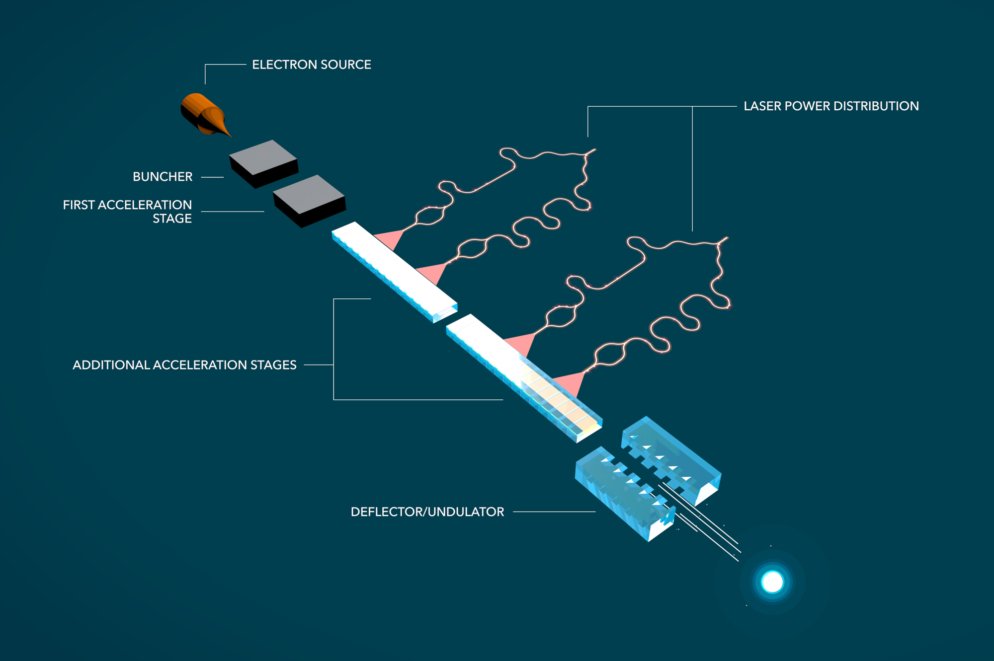
This diagram shows one possible configuration for the shoebox-sized particle accelerator prototype. The Stanford-led team will have to figure out the best way to distribute laser power among the the glass accelerating chips, generate and steer the electrons, shrink the diameter of the electron beam 1,000-fold, and a host of other technical details. (credit: SLAC National Accelerator Laboratory)

In the SLAC/Stanford experiments, published in Nature, electrons were first accelerated to nearly light speed in a SLAC accelerator test facility.  At this point they were going about as fast as they can go, and any additional acceleration would boost their energy, not their speed.

The speeding electrons then entered a chip made of silica glass and traveled through a microscopic tunnel that had tiny ridges carved into its walls. Laser light shining on the chip interacted with those ridges and produced an electrical field that boosted the energy of the passing electrons.

LCLS, the world’s first hard X-ray free-electron laser, pushes science to new extremes with ultrabright, ultrashort pulses that capture atomic-scale snapshots in quadrillionths of a second. These images can reveal never-before-seen structures and properties in matter, and can be compiled to make movies of molecules in motion. (credit: SLAC)

In the experiments, the chip achieved an acceleration gradient, or energy boost over a given distance, roughly 10 times higher than the SLAC linear accelerator can provide. At full potential, this means the 2-mile-long Linac Coherent Light Source could be replaced with a series of accelerator chips 100 meters long — roughly the length of a football field.

In a parallel approach, experiments led by Peter Hommelhoff of FAU and published in Physical Review Letters demonstrated that a laser could also be used to accelerate lower-energy electrons that had not first been boosted to nearly light speed. Both results taken together open the door to a compact particle accelerator.

Challenges
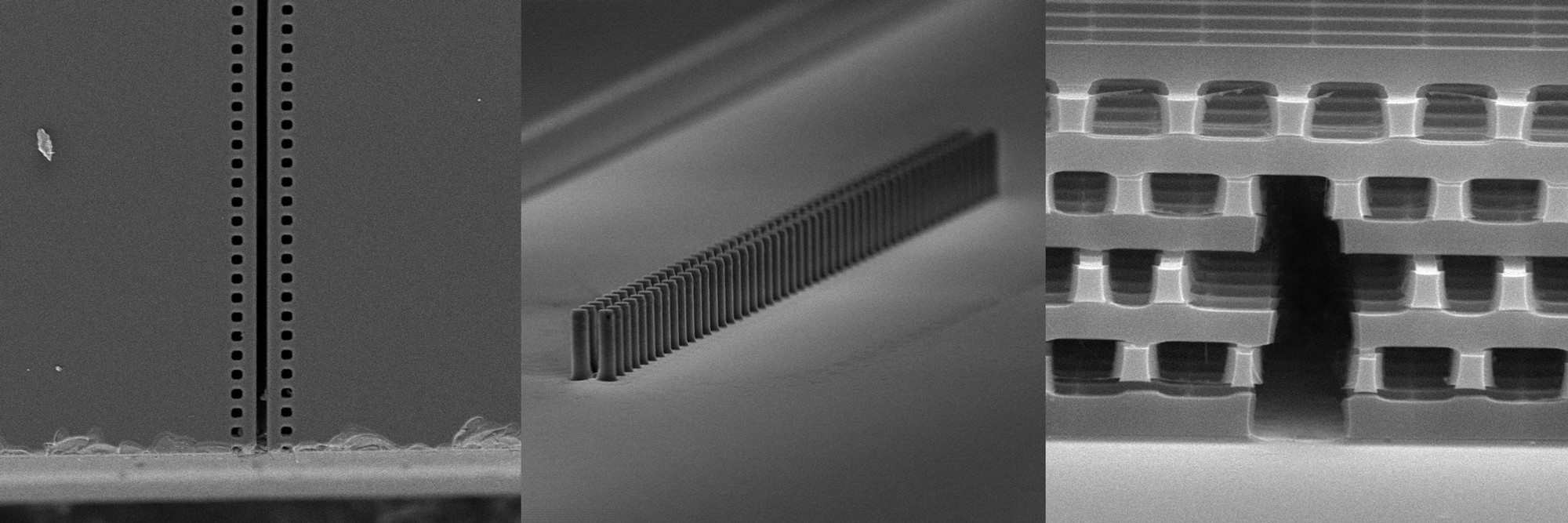
These microscopic images show some of the accelerator-on-a-chip designs being explored by the international collaboration. In each case, laser light shining on the chip boosts the energy of electrons traveling through it. (credit: Left and middle images: Andrew Ceballos, Stanford University, Right image: Chunghun Lee, SLAC)

For the past 75 years, particle accelerators have been an essential tool for physics, chemistry, biology and medicine, leading to multiple Nobel prize-winning discoveries. But without new technology to reduce the cost and size of high-energy accelerators, progress in particle physics and structural biology could stall.

The challenges of building the prototype accelerator are substantial, the scientists said. Demonstrating that a single chip works was an important step; now they must work out the optimal chip design and the best way to generate and steer electrons, distribute laser power among multiple chips and make electron beams that are 1,000 times smaller in diameter to go through the microscopic chip tunnels, among a host of other technical details.

The Stanford-led collaboration includes world-renowned experts in accelerator physics, laser physics, nanophotonics and nanofabrication. SLAC and two other national laboratories — Deutsches Elektronen-Synchrotron (DESY) in Germany and Paul Scherrer Institute in Switzerland — will contribute expertise and make their facilities available for experiments. In addition to FAU, five other universities are involved in the effort:  University of California, Los Angeles, Purdue University, University of Hamburg, the Swiss Federal Institute of Technology in Lausanne (EPFL) and Technical University of Darmstadt.


Abstract of Demonstration of electron acceleration in a laser-driven dielectric microstructure

The enormous size and cost of current state-of-the-art accelerators based on conventional radio-frequency technology has spawned great interest in the development of new acceleration concepts that are more compact and economical. Micro-fabricated dielectric laser accelerators (DLAs) are an attractive approach, because such dielectric microstructures can support accelerating fields one to two orders of magnitude higher than can radio-frequency cavity-based accelerators. DLAs use commercial lasers as a power source, which are smaller and less expensive than the radio-frequency klystrons that power today’s accelerators. In addition, DLAs are fabricated via low-cost, lithographic techniques that can be used for mass production. However, despite several DLA structures having been proposed recently, no successful demonstration of acceleration in these structures has so far been shown. Here we report high-gradient (beyond 250 MeV m−1) acceleration of electrons in a DLA. Relativistic (60-MeV) electrons are energy-modulated over 563 ± 104 optical periods of a fused silica grating structure, powered by a 800-nm-wavelength mode-locked Ti:sapphire laser. The observed results are in agreement with analytical models and electrodynamic simulations. By comparison, conventional modern linear accelerators operate at gradients of 10–30 MeV m−1, and the first linear radio-frequency cavity accelerator was ten radio-frequency periods (one metre) long with a gradient of approximately 1.6 MeV m−1. Our results set the stage for the development of future multi-staged DLA devices composed of integrated on-chip systems. This would enable compact table-top accelerators on the MeV–GeV (106–109 eV) scale for security scanners and medical therapy, university-scale X-ray light sources for biological and materials research, and portable medical imaging devices, and would substantially reduce the size and cost of a future collider on the multi-TeV (1012 eV) scale.

Multi-layer nanoparticles glow when exposed to invisible near-infrared light

An artist’s rendering shows the layers of a new, onion-like nanoparticle whose specially crafted layers enable it to efficiently convert invisible near-infrared light to higher-energy blue and UV light (credit: Kaiheng Wei)

A new onion-like nanoparticle developed at the State University of New York University at Buffalo could open new frontiers in biomaging, solar-energy harvesting, and light-based security techniques.

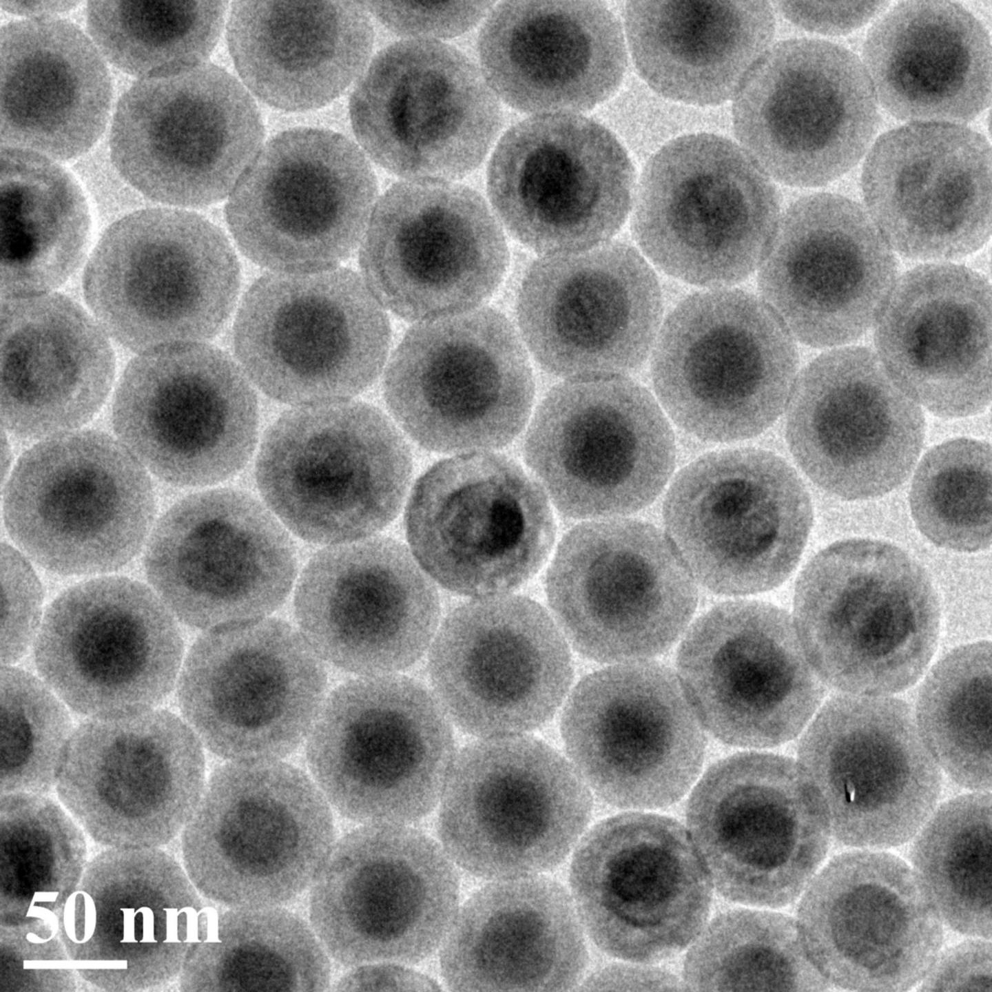
The particle’s innovation lies in its layers: a coating of organic dye, a neodymium-containing shell, and a core that incorporates ytterbium and thulium. Together, these strata convert invisible near-infrared light to higher energy blue and UV light with record-high efficiency.

A transmission electron microscopy image of the new nanoparticles, which convert invisible near-infrared light to higher-energy blue and UV light with high efficiency. Each particle is about 50 nanometers in diameter. (credit: Institute for Lasers, Photonics and Biophotonics, University at Buffalo)

Light-emitting nanoparticles placed by a surgeon inside the body could provide high-contrast images of areas of interest. Nanoparticle-infused inks could also be incorporated into currency designs using ink that is invisible to the naked eye but glows blue when hit by a low-energy near-infrared laser pulse — which would very difficult for counterfeiters to reproduce.

The researchers say the nanoparticle is about 100 times more efficient at “upconverting” [increasing the frequency of] light than similar nanoparticles.

Peeling back the layers

Energy-cascaded upconversion (credit: Guanying Chen et al./Nano Letters)

Converting low-energy light to light of higher energies is difficult to do. It involves capturing two or photons from a low-energy light source, and combining their energy to form a single, higher-energy photon. Each of the three layers of this onionesque nanoparticle fulfills a unique function:

  • The outermost layer is a coating of organic dye. This dye is adept at absorbing photons from low-energy near-infrared light sources. It acts as an “antenna” for the nanoparticle, harvesting light and transferring energy inside, Ohulchanskyy says.
  • The next layer is a neodymium-containing shell. This layer acts as a bridge, transferring energy from the dye to the particle’s light-emitting core.*
  • Inside the light-emitting core, ytterbium and thulium ions work in concert. The ytterbium ions draw energy into the core and pass the energy on to the thulium ions, which have special properties that enable them to absorb the energy of three, four or five photons at once, and then emit a single higher-energy photon of blue and UV light.

The research was published online in Nano Letters on Oct. 21. It was led by the Institute for Lasers, Photonics, and Biophotonics at UB, and the Harbin Institute of Technology in China, with contributions from the Royal Institute of Technology in Sweden, Tomsk State University in Russia, and the University of Massachusetts Medical School.

* The neodymium-containing layer is necessary for transferring energy efficiently from dye to core. When molecules or ions in a material absorb a photon, they enter an “excited” state from which they can transfer energy to other molecules or ions. The most efficient transfer occurs between molecules or ions whose excited states require a similar amount of energy to obtain, but the dye and ytterbium ions have excited states with very different energies. So the team added neodymium — whose excited state is in between that of the dye and thulium’s — to act as a bridge between the two, creating a “staircase” for the energy to travel down to reach emitting thulium ions.


Abstract of Energy-Cascaded Upconversion in an Organic Dye-Sensitized Core/Shell Fluoride Nanocrystal

Lanthanide-doped upconversion nanoparticles hold promises for bioimaging, solar cells, and volumetric displays. However, their emission brightness and excitation wavelength range are limited by the weak and narrowband absorption of lanthanide ions. Here, we introduce a concept of multistep cascade energy transfer, from broadly infrared-harvesting organic dyes to sensitizer ions in the shell of an epitaxially designed core/shell inorganic nanostructure, with a sequential nonradiative energy transfer to upconverting ion pairs in the core. We show that this concept, when implemented in a core–shell architecture with suppressed surface-related luminescence quenching, yields multiphoton (three-, four-, and five-photon) upconversion quantum efficiency as high as 19% (upconversion energy conversion efficiency of 9.3%, upconversion quantum yield of 4.8%), which is about ∼100 times higher than typically reported efficiency of upconversion at 800 nm in lanthanide-based nanostructures, along with a broad spectral range (over 150 nm) of infrared excitation and a large absorption cross-section of 1.47 × 10⁻¹⁴ cm2 per single nanoparticle. These features enable unprecedented three-photon upconversion (visible by naked eye as blue light) of an incoherent infrared light excitation with a power density comparable to that of solar irradiation at the Earth surface, having implications for broad applications of these organic–inorganic core/shell nanostructures with energy-cascaded upconversion.

New technology senses colors in the infrared spectrum

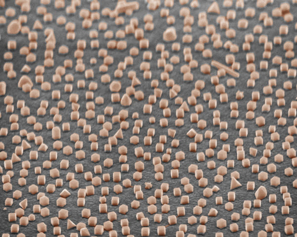
A scanning electron microscope image showing a surface coated with silver nanocubes on a gold surface to image near-infrared light (credit: Maiken Mikkelsen and Gleb Akselrod, Duke University)

Duke University scientists have invented a technology that can identify and image different wavelengths of the infrared spectrum.

The fabrication technique for the system is easily scalable, can be applied to any surface geometry, and costs much less than current light-absorption technologies, according to the researchers. Once adopted, the technique would allow advanced infrared imaging systems to be produced faster and cheaper than today’s counterparts and with higher sensitivity.

The visible-light spectrum, with wavelengths of about 400 to 700 nanometers. The near-infrared region starts just above the red region. (credit: Wikipedia)

Using nanoparticles to absorb specific wavelengths

The technology relies on a physics phenomenon called plasmonics to absorb or reflect specific wavelengths, similar to how stained-glass windows are created (see “Crafting color coatings from nanometer-thick layers of gold and germanium“).

A curved object covered with a coating of 100-nanometer silver cubes that absorbs all red light, leaving the object with a green tint (credit: Maiken Mikkelsen and Gleb Akselrod, Duke University)

The researchers first coat a surface with a thin film of gold through a common process like evaporation. They then put down a few-nanometer-thin layer of polymer, followed by a coating of silver cubes, each one about 100 nanometers (billionths of a meter) in size.

When light strikes the new engineered surface, a specific color gets trapped on the surface of the nanocubes in packets of energy called plasmons, and eventually dissipates into heat. By controlling the thickness of the polymer film and the size and number of silver nanocubes, the coating can be tuned to absorb different wavelengths of light from the visible spectrum (at 650 nm) to the near infrared (up to 1420 nm).

“By borrowing well-known techniques from chemistry and employing them in new ways, we were able to obtain significantly better resolution than with a million-dollar state-of-the-art electron beam lithography system,” said Maiken H. Mikkelsen, the Nortel Networks Assistant Professor of Electrical & Computer Engineering and Physics at Duke University. “This allowed us to create a coating that can fine-tune the absorption spectra with a level of control that hasn’t been possible previously.”

Coating photodetectors to absorb specific wavelengths of near-infrared light would allow novel and cheap cameras to be made that could capture and discriminate different wavelengths.

A better “tricorder” camera

The researchers note in the paper that “plasmonic resonances could be moved deeper into the infrared spectrum by using larger colloidal nanoparticles with larger metallic facets,” including wavelengths in the thermal infrared spectrum.

Colors shown on current thermal infrared-imaging displays are actually based on a simple pseudocolor scheme in which the color displayed is arbitrary — it only represents the amount of thermal radiation (infrared light) that the camera captures, not its wavelength in the electromagnetic spectrum.

That means the technology could be used in a variety of other applications, such as masking the heat signatures of objects such as stealth aircraft (see “Plasmonic cloaking“) or creating a sophisticated “tricorder” camera that shows a person’s temperature at different mid-infrared (thermal) wavelengths (see “Graphene could take night-vision thermal imagers beyond ‘Predator’“).

The study was published online Nov. 9 in the journal Advanced Materials and was supported by the Air Force Office of Scientific Research.


Abstract of Large-Area Metasurface Perfect Absorbers from Visible to Near-Infrared

An absorptive metasurface based on film-coupled colloidal silver nanocubes is demonstrated. The metasurfaces are fabricated using simple dip-coating methods and can be deposited over large areas and on arbitrarily shaped objects. The surfaces show nearly complete absorption, good off-angle performance, and the resonance can be tuned from the visible to the near-infrared.

‘Golden window’ wavelength range for optimal deep-brain near-infrared imaging determined

Rayleigh scattering causes the reddening of the sun at sunset — an example of how longer wavelengths (yellow and red compared to blue in blue sky) penetrate matter (dust at sunset) better. (credit: Wikipedia/CC)

Researchers at The City College of New York (CCNY) have determined the optimal wavelengths for bioimaging of the brain at longer near-infrared wavelengths, which permit deeper imaging.

Near-infrared (NIR) radiation has been used for one- and two-photon fluorescence imaging at near-infrared wavelengths of 650–950 nm (nanometers) for deep brain imaging, but it is limited in penetration depth. (The CCNY researchers dubbed this Window I, also known as the therapeutic window.)

Longer infrared wavelengths penetrate deeper but are limited by Rayleigh and Mie scattering, which blur images, and absorption, which reduces the number of available photons (brightness). These limitations are based on the lack of suitable CMOS semiconductor imaging detectors or femtosecond laser sources.

The new CCNY study, led by biomedical engineer Lingyan Shi, studied three new optical windows in the near-infrared (NIR) region, in addition to Window I, for high-resolution deep brain imaging.

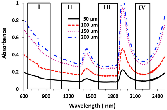
Their study built on a prior CCNY study* in 2014 using detectors based on indium gallium arsenide (GaAs) or indium antimonide-(InSb) and a femtosecond excitation source of IMRA fiber laser** to image rat brain tissue in window II (1,100– 1,350 nm), window III (1,600–1,870 nm), and window IV (1600 nm to 1870 nm, centered at 2,200 nm).

Absorbance for four windows at four brain-tissue thicknesses (credit: Lingyan Shi et al./J. Biophotonics)

The new CCNY research investigated the optimal wavelength band and optical properties of brain tissue with NIR, including the total attenuation coefficient (μt), absorption coefficient (μa), reduced scattering coefficient (μ0 s), and the scattering anisotropy coefficient (g) in these optical windows. The purpose of the study was to determine an optimal optical window in NIR in the 650 nm to 2500 nm range to reduce scattering, achieve optimal absorption, and reduce noise for deep-brain tissue imaging.

Golden Window: optimal wavelength range

Peak transmittance T (%) measured with each optical window for brain tissues with four thicknesses. Window III had the highest transmittance percentage for each of the thicknesses, followed by windows II and IV. (credit: Lingyan Shi et al./J. Biophotonics)

The researchers found that the “Golden Window” (1600 nm to 1870 nm) is an optimal wavelength range for light penetration in brain tissue, followed by Windows II and IV.

“This is a first for brain imaging and proved theoretically and experimentally that deep imaging of the brain is possible using light at longer wavelengths. It demonstrates these windows’ potential for deeper brain tissue imaging due to the reduction of scattering that causes blurring,” said Shi, a research associate in City College’s Institute for Ultrafast Spectroscopy and Lasers, and the biology department.

Published by the Journal of Biophotonics, her study sheds light on the development of the next generation of microscopy imaging technique, in which the “Golden Window” may be utilized for high-resolution deeper brain imaging. The next step in the research is in vivo imaging in mice using Golden Window wavelength light.

Shi’s team included Distinguished Professor of Physics Robert R. Alfano and Adrian Rodriguez-Contreras, an assistant professor of biology. Shi earned a Ph.D. in biomedical engineering from CCNY’s Grove School of Engineering in 2014.

* L. A. Sordillo, Y. Pu, S. Pratavieira, Y. Budansky, and R. R. Alfano, J. Biomed. Opt. 19, 056004 (2014) [link].

** Excitation wavelength 1680 nm, power > 200 mW, pulse width 100 fs, and 50 MHz repetition rate.


Abstract of Transmission in near-infrared optical windows for deep brain imaging

Near-infrared (NIR) radiation has been employed using one- and two-photon excitation of fluorescence imaging at wavelengths 650–950 nm (optical window I) for deep brain imaging; however, longer wavelengths in NIR have been overlooked due to a lack of suitable NIR-low band gap semiconductor imaging detectors and/or femtosecond laser sources. This research introduces three new optical windows in NIR and demonstrates their potential for deep brain tissue imaging. The transmittances are measured in rat brain tissue in the second (II, 1,100–1,350 nm), third (III, 1,600–1,870 nm), and fourth (IV, centered at 2,200 nm) NIR optical tissue windows. The relationship between transmission and tissue thickness is measured and compared with the theory. Due to a reduction in scattering and minimal absorption, window III is shown to be the best for deep brain imaging, and windows II and IV show similar but better potential for deep imaging than window I.

New dimension to high-temperature superconductivity discovered

In this artistic rendering, a magnetic pulse (right) and X-ray laser light (left) converge on a high-temperature superconductor to study the behavior of its electrons (credit: SLAC National Accelerator Laboratory)

The dream to push the operating temperature for superconductors to room temperature — leading to future advances in computing, electronics and power grid technologies — has just become more real.

A team led by scientists at the Department of Energy’s SLAC National Accelerator Laboratory has combined powerful magnetic pulses with some of the brightest X-rays on the planet, discovering a surprising 3-D arrangement of a material’s electrons that appears closely linked to high-temperature superconductivity.

The scientists say this unexpected twist marks an important milestone in the 30-year journey to better understand how materials known as high-temperature superconductors conduct electricity with no resistance at temperatures hundreds of degrees Fahrenheit above those of conventional metal superconductors (but still hundreds of degrees below freezing). The study was published today (Nov. 5) in Science.

There are already many uses for standard low-temperature superconducting technology, from MRI machines that diagnose brain tumors to a prototype levitating train, the CERN particle collider that enabled the Nobel Prize-winning discovery of the Higgs boson, and ultrasensitive detectors used to hunt for dark matter — the invisible constituent believed to make up most of the mass of the universe.

‘Totally unexpected’ physics

“This was totally unexpected, and also very exciting. This experiment has identified a new ingredient to consider in this field of study. Nobody had seen this 3-D picture before,” said Jun-Sik Lee, a SLAC staff scientist and one of the leaders of the experiment conducted at SLAC’s Linac Coherent Light Source (LCLS) X-ray laser.  (A planned upgrade to the LCLS, known as LCLS-II, will include a superconducting particle accelerator.) “This is an important step in understanding the physics of high-temperature superconductors.”

The 3-D effect that scientists observed in the LCLS experiment, which occurs in a superconducting material known as YBCO (yttrium barium copper oxide), is a newly discovered type of “charge density wave.” This wave does not have the oscillating motion of a light wave or a sound wave; it describes a static, ordered arrangement of clumps of electrons in a superconducting material. Its coexistence with superconductivity is perplexing to researchers because it seems to conflict with the freely moving electron pairs that define superconductivity.

The 2-D version of this wave was first seen in 2012 and has been studied extensively. The LCLS experiment revealed a separate 3-D version that appears stronger than the 2-D form and closely tied to both the 2-D behavior and the material’s superconductivity.

The experiment was several years in the making and required international expertise to prepare the specialized samples and construct a powerful customized magnet that produced magnetic pulses compressed to thousandths of a second. Each pulse was 10-20 times stronger than those from the magnets in a typical medical MRI machine.

A powerful blend of magnetism and light

This custom-made magnet was used in an experiment at SLAC’s Linac Coherent Light Source X-ray laser to study an effect known as a charge density wave. (credit: Jun-Sik Lee)

Those short but intense magnetic pulses suppressed the superconductivity of the YBCO samples and provided a clearer view of the charge density wave effects. They were immediately followed at precisely timed intervals by ultra-bright LCLS X-ray laser pulses, which allowed scientists to measure the wave effects.

“This experiment is a completely new way of using LCLS that opens up the door for a whole new class of future experiments,” said Mike Dunne, LCLS director.*

“I’ve been excited about this experiment for a long time,” said Steven Kivelson, a Stanford University physics professor who contributed to the study and has researched high-temperature superconductors since 1987.

Kivelson said the experiment sets very clear boundaries on the temperature and strength of the magnetic field at which the newly observed 3-D effect emerges. “There is nothing vague about this,” he said. “You can now make a definitive statement: In this material, a new phase exists.”

The experiment also adds weight to the growing evidence that charge density waves and superconductivity “can be thought of as two sides of the same coin,” he added.

A view of the X-ray Correlation Spectroscopy experimental station at SLAC’s Linac Coherent Light Source (LCLS) X-ray laser. This station was used for an experiment studying an effect in a superconducting material. (credit: SLAC National Accelerator Laboratory)

A more complete map

But it is also clear that YBCO is incredibly complex, and a more complete map of all of its properties is required to reach any conclusions about what matters most to its superconductivity, said Simon Gerber of SIMES and Hoyoung Jang of SSRL, the lead authors of the study.

Follow-up experiments are needed to provide a detailed visualization of the 3-D effect, and to learn whether the effect is universal across all types of high-temperature superconductors, said SLAC staff scientist and SIMES investigator Wei-Sheng Lee, who co-led the study with Jun-Sik Lee of SSRL and Diling Zhu of LCLS. “The properties of this material are much richer than we thought,” Lee said.

“We continue to make new and surprising observations as we develop new experimental tools,” Zhu added.

* Researchers conducted many preparatory experiments at SLAC’s Stanford Synchrotron Radiation Lightsource (SSRL), which also produces X-rays for research. LCLS and SSRL are DOE Office of Science User Facilities. Scientists from SIMES, the Stanford Institute for Materials and Energy Sciences at SLAC, and SSRL and LCLS were a part of the study.

Abstract of Three-Dimensional Charge Density Wave Order in YBa2Cu3O6.67 at High Magnetic Fields

Charge density wave (CDW) correlations have been shown to universally exist in cuprate superconductors. However, their nature at high fields inferred from nuclear magnetic resonance is distinct from that measured by x-ray scattering at zero and low fields. Here we combine a pulsed magnet with an x-ray free electron laser to characterize the CDW in YBa2Cu3O6.67 via x-ray scattering in fields up to 28 Tesla. While the zero-field CDW order, which develops below T ~ 150 K, is essentially two-dimensional, at lower temperature and beyond 15 Tesla, another three-dimensionally ordered CDW emerges. The field-induced CDW appears around the zero-field superconducting transition temperature; in contrast, the incommensurate in-plane ordering vector is field-independent. This implies that the two forms of CDW and high-temperature superconductivity are intimately linked.

China plans world’s largest supercollider

China’s Circular Electron Positron Collider (CEPC) is expected to be at least twice the size of the world’s current leading collider, the Large Hadron Collider at CERN, partially shown here (credit: Maximilien Brice/CERN)

Chinese scientists are completing plans for the Circular Electron Positron Collider (CEPC), a supergiant particle collider. With a circumference of 80 kilometers (50 miles) when built, it will be at least twice the size of the world’s current leading collider, the Large Hadron Collider at CERN, outside Geneva, according to the Institute of High Energy Physics in Beijing. Work on the collider is expect to start in 2020.

The collider complex is initially designed to smash together electrons and their anti-matter counterparts, and later more massive protons, at velocities approaching the speed of light. This process hopes to recreate, inside the accelerator, the hyper-energy conditions that dominated following the Big Bang.

Physicists aim to explore the origins of matter, energy, and space-time. China says its collider will ultimately be able to reach higher energy levels than CERN; this might help physicists discover a new range of particles beyond those already charted in the Standard Model of Particle Physics.

According to Professor Nima Arkani-Hamed, a scholar at Princeton’s Institute for Advanced Study, a perfect circle-shaped city, hosting the globe’s leaders in experimental particle physics, new-technology firms and other future-oriented scholars and designers, could be created inside the massive Chinese collider complex. The complex would also host a multipurpose science-technology campus aimed at conducting secondary and supplemental science experiments.

Within the same 80km tunnel, the collider complex plans to be divided between two different super colliders. The Circular Electron Positron Collider (CEPC) is designed to study the Higgs boson and how it decays following a collision between electrons and anti-electrons. The Super Proton Proton Collider (SPPC) will be used to study the super-speed collisions of protons.

New quadrupole magnets, which focus particle beams before collisions, are one of the key technologies for the High-Luminosity LHC. (credit: CERN)

Last week, more than 230 scientists and engineers from around the world met at CERN to discuss the High-Luminosity LHC — a major upgrade to the Large Hadron Collider (LHC) that “will increase its discovery potential from 2025,” according to the CERN website.

Massive supercomputer simulation models universe from near birth until today

Galaxies have halos surrounding them, which may be composed of both dark and regular matter. This image shows a substructure within a halo in the Q Continuum simulation, with “subhalos” marked in different colors. (credit: Heitmann et al.)

The Q Continuum simulation, one of the largest cosmological simulations ever performed, has modeled the evolution of the universe from just 50 million years after the Big Bang to the present day.

DOE’s Argonne National Laborator led the simulation on the Titan supercomputer at DOE’s Oak Ridge National Laboratory.

Over the course of 13.8 billion years, the matter in the universe clumped together to form galaxies, stars, and planets. These kinds of simulations help scientists understand dark energy (a form of energy that affects the expansion rate of the universe) and the distribution of galaxies, composed of ordinary matter and mysterious dark matter.

This series run on the Titan supercomputer simulates the evolution of the universe. The images give an impression of the detail in the matter distribution in the simulation. At first, the matter is very uniform, but over time, gravity acts on the dark matter, which begins to clump more and more, and in the clumps, galaxies form. (credit: Heitmann et. al.)

Intensive sky surveys with powerful telescopes, like the Sloan Digital Sky Survey and the new, more detailed Dark Energy Survey, show scientists where galaxies and stars were when their light was first emitted. And surveys of the Cosmic Microwave Background (light remaining from when the universe was only 300,000 years old) show us how the universe began — “very uniform, with matter clumping together over time,” said Katrin Heitmann, an Argonne physicist who led the simulation.

The simulation fills in the temporal gap to show how the universe might have evolved in between: “Gravity acts on the dark matter, which begins to clump more and more, and in the clumps, galaxies form,” said Heitmann.

The Q Continuum simulation involved half a trillion particles — dividing the universe up into cubes with sides 100,000 kilometers long. This makes it one of the largest cosmology simulations at such high resolution. It ran using more than 90 percent of the supercomputer.

“This is a very rich simulation,” Heitmann said. “We can use this data to look at why galaxies clump this way, as well as the fundamental physics of structure formation itself.”

Analysis has already begun on the two and a half petabytes of data that were generated, and will continue for several years, she said. Scientists can pull information on such astrophysical phenomena as strong lensing, weak lensing shear, cluster lensing, and galaxy-galaxy lensing.


Abstract of The Q Continuum simulation: Harnessing the power of GPU accelerated supercomputers

Modeling large-scale sky survey observations is a key driver for the continuing development of high-resolution, large-volume, cosmological simulations. We report the first results from the “Q Continuum” cosmological N-body simulation run carried out on the GPU-accelerated supercomputer Titan. The simulation encompasses a volume of (1300 Mpc)3 and evolves more than half a trillion particles, leading to a particle mass resolution of ${m}_{{rm{p}}}simeq 1.5cdot {10}^{8};$ ${M}_{odot }$. At this mass resolution, the Q Continuum run is currently the largest cosmology simulation available. It enables the construction of detailed synthetic sky catalogs, encompassing different modeling methodologies, including semi-analytic modeling and sub-halo abundance matching in a large, cosmological volume. Here we describe the simulation and outputs in detail and present first results for a range of cosmological statistics, such as mass power spectra, halo mass functions, and halo mass-concentration relations for different epochs. We also provide details on challenges connected to running a simulation on almost 90% of Titan, one of the fastest supercomputers in the world, including our usage of Titan’s GPU accelerators.
Holographic sonic tractor beam lifts and moves objects using soundwaves

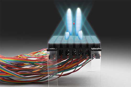
Holograms (3-D light fields) can be projected from a 2-dimensional surface to control objects. (credit: Asier Marzo, Bruce Drinkwater and Sriram Subramanian)

British researchers have built a working Star-Trek-style “tractor beam” — a device that can attract or repel one object to another from a distance. It uses high-amplitude soundwaves to generate an acoustic hologram that can grasp and move small objects.

The technique, published in an open-access paper in Nature Communications October 27, has a wide range of potential applications, the researchers say. A sonic production line could transport delicate objects and assemble them, all without physical contact. Or a miniature version could grip and transport drug capsules or microsurgical instruments through living tissue.

The device was developed at the Universities of Sussex and Bristol in collaboration with Ultrahaptics.


University of Sussex | Levitation using sound waves

The researchers used an array of 64 miniature loudspeakers. The whole system consumes just 9 Watts of power, used to create high-pitched (40Khz), high-intensity sound waves to levitate a spherical bead 4mm in diameter made of expanded polystyrene.

The tractor beam works by surrounding the object with high-intensity sound to create a force field that keeps the objects in place. By carefully controlling the output of the loudspeakers, the object can be held in place, moved, or rotated.

Three different shapes of acoustic force fields work as tractor beams: an acoustic force field that resembles a pair of fingers or tweezers; an acoustic vortex, the objects becoming trapped at the core; and a high-intensity “cage” that surrounds the objects and holds them in place from all directions.

Previous attempts surrounded the object with loudspeakers, which limits the extent of movement and restricts many applications. Last year, the University of Dundee presented the concept of a tractor beam, but no objects were held in the ray.

The team is now designing different variations of this system. A bigger version aims at levitating a soccer ball from 10 meters away and a smaller version aims at manipulating particles inside the human body.


Asier Marzo, Matt Sutton, Bruce Drinkwater and Sriram Subramanian | Acoustic holograms are projected from a flat surface and contrary to traditional holograms, they exert considerable forces on the objects contained within. The acoustic holograms can be updated in real time to translate, rotate and combine levitated particles enabling unprecedented contactless manipulators such as tractor beams.


Abstract of Holographic acoustic elements for manipulation of levitated objects

Sound can levitate objects of different sizes and materials through air, water and tissue. This allows us to manipulate cells, liquids, compounds or living things without touching or contaminating them. However, acoustic levitation has required the targets to be enclosed with acoustic elements or had limited maneuverability. Here we optimize the phases used to drive an ultrasonic phased array and show that acoustic levitation can be employed to translate, rotate and manipulate particles using even a single-sided emitter. Furthermore, we introduce the holographic acoustic elements framework that permits the rapid generation of traps and provides a bridge between optical and acoustical trapping. Acoustic structures shaped as tweezers, twisters or bottles emerge as the optimum mechanisms for tractor beams or containerless transportation. Single-beam levitation could manipulate particles inside our body for applications in targeted drug delivery or acoustically controlled micro-machines that do not interfere with magnetic resonance imaging.

Mass extinctions linked to comet and asteroid showers

Mass extinctions occurring over the past 260 million years were likely caused by comet and asteroid showers, a new study concludes. An artist’s illustration of a major asteroid impact on Earth. (credit: NASA/Don Davis)

Mass extinctions occurring over the past 260 million years were likely caused by comet and asteroid showers, scientists conclude in a new study published in an open-access paper in Monthly Notices of the Royal Astronomical Society.

For more than 30 years, scientists have argued about a controversial hypothesis relating to periodic mass extinctions and impact craters — caused by comet and asteroid showers — on Earth.

In their MNRAS paper, Michael Rampino, a New York University geologist, and Ken Caldeira, a scientist in the Carnegie Institution’s Department of Global Ecology, offer new support linking the age of these craters with recurring mass extinctions of life every 26 million years, including the demise of dinosaurs.

This cycle has been linked to periodic motion of the sun and planets through the dense mid-plane of our galaxy. Scientists have theorized that gravitational perturbations of the distant Oort comet cloud that surrounds the sun lead to periodic comet showers in the inner solar system, where some comets strike the Earth.

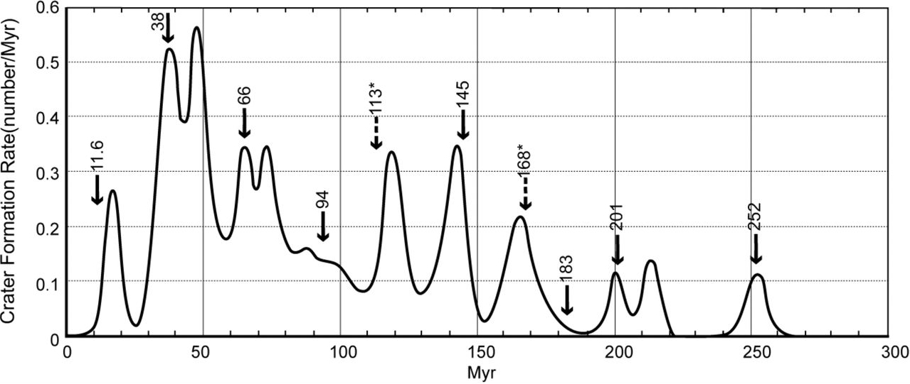
Crater formation rate per million years, with eight significant extinction events shown with solid arrows and two potential extinction events shown with broken arrows (credit: Michael R. Rampino and Ken Caldeira/MNRAS)

To test their hypothesis, Rampino and Caldeira performed time-series analyses of impacts and extinctions using newly available data offering more accurate age estimates. “The correlation between the formation of these impacts and extinction events over the past 260 million years is striking and suggests a cause-and-effect relationship,” says Rampino.

The sinkholes clustered around the trough of the Chicxulub crater suggest a prehistoric oceanic basin in the depression left by the impact. (credit: NASA)

One of the craters considered in the study is the large (180 km diameter) Chicxulub impact structure in the Yucatan, which dates at about 65 million years ago — the time of a great mass extinction that included the dinosaurs. And five out of the six largest impact craters of the last 260 million years on earth correlate with mass extinction events.


Abstract of Periodic impact cratering and extinction events over the last 260 million years

The claims of periodicity in impact cratering and biological extinction events are controversial. A newly revised record of dated impact craters has been analyzed for periodicity, and compared with the record of extinctions over the past 260 Myr. A digital circular spectral analysis of 37 crater ages (ranging in age from 15 to 254 Myr ago) yielded evidence for a significant 25.8 ± 0.6 Myr cycle. Using the same method, we found a significant 27.0 ± 0.7 Myr cycle in the dates of the eight recognized marine extinction events over the same period. The cycles detected in impacts and extinctions have a similar phase. The impact crater dataset shows 11 apparent peaks in the last 260 Myr, at least 5 of which correlate closely with significant extinction peaks. These results suggest that the hypothesis of periodic impacts and extinction events is still viable.

Largest astronomical image to date contains 46 billion pixels

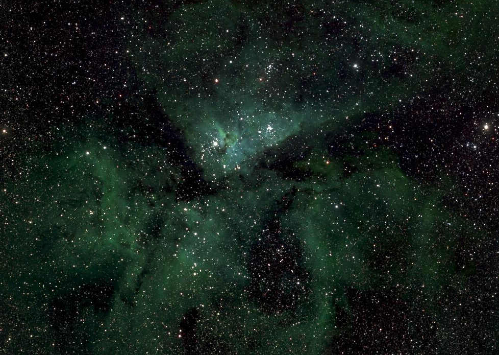
A small section of the Milky Way photo showing the star Eta Carinae (credit: Lehrstuhl für Astrophysik/RUB)

Astronomers at the Ruhr-Universität Bochum in Germany have compiled the largest astronomical image to date: a picture of the Milky Way containing 46 billion pixels, viewable here (you can enter an object name, such as “Eta Carinae,” in the lower-left box).

The image was generated over a period of five years of astronomical observations by two telescopes at Bochum’s university observatory in the Atacama Desert in Chile. It only includes objects with variable brightness, which includes stars with a planet passing in front. The area that the astronomers observed is so large that they had to subdivide it into 268 sections.

False color image of the field containing the Galactic Center (credit: M. Haas et al./Astronomical Notes)


Abstract of The Bochum survey of the southern Galactic disk: I. Survey design and first results on 50 square degrees monitored in 2011

We are monitoring a 6° wide stripe along the southern Galactic disk simultaneously in the r and i bands, using a robotic 15-cm twin telescope of the Universitätsternwarte Bochum near Cerro Armazones in Chile. Utilising the telescope’s 2.7° field of view, the survey aims at observing a mosaic of 268 fields once per month and to monitor dedicated fields once per night. The survey reaches a sensitivity from 10m down to 18m (AB system), with a completeness limit of r ∼ 15.5m and i ∼ 14.5m which – due to the instrumental pixel size of 2.″4 – refers to stars separated by >3″. This brightness range is ideally suited to examine the intermediately bright stellar population supposed to be saturated in deep variability surveys with large telescopes. To connect to deep surveys or to explore faint long term variables, coadded images of several nights reach a depth of ∼ 20m. The astrometric accuracy is better than 1″, as determined from the overlap of neighbouring fields. We describe the survey design, the data properties and our procedures to derive the light curves and to extract variable stars. We present a list of ∼2200 variable stars identified in 50 square degrees with 50-80 observations between May and October 2011. For bright stars the variability amplitude A reaches down to A ∼ 0.05m, while at the faint end variations of A > 1m are detected. About 200 stars were known tobe variable, and their amplitudes and periods – as far as determinable from our six month monitoring – agree with literature values, demonstrating the performance of the Bochum Galactic Disk Survey (© 2012 WILEY-VCH Verlag GmbH & Co. KGaA, Weinheim)


Abstract of The Bochum Survey of the Southern Galactic Disk: II. Follow-up measurements and multi-filter photometry for 1323 square degrees monitored in 2010 – 2015

This paper is the second in a series describing the southern Galactic Disk Survey (GDS) performed at the Universitätssternwarte Bochum near Cerro Armazones in Chile. Haas et al. (2012, Paper I) presented the survey design and the characteristics of the observations and data. They identified ∼2200 variable stars in an area of 50 square degrees with more than 50 observations in 2011. Here we present the first complete version of the GDS covering all 268 fields with 1323 square degrees along the Galactic disk including revised data from Paper I. The individual fields were observed up to 272 times and comprise a maximum time span between September 2010 and May 2015. We detect a total of 64 151 variable sources, which are presented in a catalog including some of their properties and their light curves. A comparison with the International Variable Star Index (VSX) and All Sky Automated Survey (ASAS) indicates that 56794 of these sources are previously unknown variables. Furthermore, we present UBVr ′, i ′, z ′ photometry for all sources within the GDS, resulting in a new multi-color catalog of nearly 16×106 sources detected in at least one filter. Both the GDS and the near-infrared VISTA Variables in the Via Lactea survey (VVV) complement each other in the overlap area of about 300 square degrees enabling future comparison studies. (© 2015 WILEY-VCH Verlag GmbH & Co. KGaA, Weinheim)