How to program DNA like we do computers

A programmable chemical oscillator made from DNA (credit: Ella Maru Studio and Cody Geary)

Researchers at The University of Texas at Austin have programmed DNA molecules to follow specific instructions to create sophisticated molecular machines that could be capable of communication, signal processing, problem-solving, decision-making, and control of motion in living cells — the kind of computation previously only possible with electronic circuits.

Future applications may include health care, advanced materials, and nanotechnology.

As a demonstration, the researchers constructed a first-of-its-kind chemical oscillator that uses only DNA components — no proteins, enzymes or other cellular components — to create a classic chemical reaction network (CRN) called a “rock-paper-scissors oscillator.” The goal was to show that DNA alone is capable of precise, complex behavior.

A systematic pipeline for programming DNA-only dynamical systems and the implementation of a chemical oscillator (credit: Niranjan Srinivas et al./Science)

Chemical oscillators have long been studied by engineers and scientists. For example, the researchers who discovered the chemical oscillator that controls the human circadian rhythm — responsible for our bodies’ day and night rhythm — earned the 2017 Nobel Prize in physiology or medicine.

“As engineers, we are very good at building sophisticated electronics, but biology uses complex chemical reactions inside cells to do many of the same kinds of things, like making decisions,” said David Soloveichik, an assistant professor in the Cockrell School’s Department of Electrical and Computer Engineering and senior author of a paper in the journal Science.

“Eventually, we want to be able to interact with the chemical circuits of a cell, or fix malfunctioning circuits or even reprogram them for greater control. But in the near term, our DNA circuits could be used to program the behavior of cell-free chemical systems that synthesize complex molecules, diagnose complex chemical signatures, and respond to their environments.”

The team’s research was conducted as part of the National Science Foundation’s (NSF) Molecular Programming Project and funded by the NSF, the Office of Naval Research, the National Institutes of Health, and the Gordon and Betty Moore Foundation.


Programming a Chemical Oscillator


Abstract of Enzyme-free nucleic acid dynamical systems

An important goal of synthetic biology is to create biochemical control systems with the desired characteristics from scratch. Srinivas et al. describe the creation of a biochemical oscillator that requires no enzymes or evolved components, but rather is implemented through DNA molecules designed to function in strand displacement cascades. Furthermore, they created a compiler that could translate a formal chemical reaction network into the necessary DNA sequences that could function together to provide a specified dynamic behavior.


Controlled by a synthetic gene circuit, self-assembling bacteria build working electronic sensors

Bacteria create a functioning 3D pressure-sensor device. A gene circuit (left) triggers the production of an engineered protein that enables pattern-forming bacteria on growth membranes (center) to assemble gold nanoparticles into a hybrid organic-inorganic dome structure whose size and shape can be controlled by altering the growth environment. In this proof-of-concept demonstration, the gold structure serves as a functioning pressure switch (right) that responds to touch. (credit: Yangxiaolu Cao et al./Nature Biotechnology)

Using a synthetic gene circuit, Duke University researchers have programmed self-assembling bacteria to build useful electronic devices — a first.

Other experiments have successfully grown materials using bacterial processes (for example, MIT engineers have coaxed bacterial cells to produce biofilms that can incorporate nonliving materials, such as gold nanoparticles and quantum dots). However, they have relied entirely on external control over where the bacteria grow and they have been limited to two dimensions.

In the new study, the researchers demonstrated the production of a composite structure by programming the cells themselves and controlling their access to nutrients, but still leaving the bacteria free to grow in three dimensions.*

As a demonstration, the bacteria were programmed to assemble into a finger-pressure sensor.

To create the pressure sensor, two identical arrays of domes were grown on a membrane (left) on two substrate surfaces. The two substrates were then sandwiched together (center) so that each dome was positioned directly above its counterpart on the other substrate. A battery was connected to the domes by copper wiring. When pressure was applied (right) to the sandwich, the domes pressed into one another, causing a deformation, resulting in an increase in conductivity, with resulting increased current (as shown the arrow in the ammeter). (credit: Yangxiaolu Cao et al./Nature Biotechnology)

Inspired by nature, but going beyond it

“This technology allows us to grow a functional device from a single cell,” said Lingchong You, the Paul Ruffin Scarborough Associate Professor of Engineering at Duke. “Fundamentally, it is no different from programming a cell to grow an entire tree.”

Nature is full of examples of life combining organic and inorganic compounds to make better materials. Mollusks grow shells consisting of calcium carbonate interlaced with a small amount of organic components, resulting in a microstructure three times tougher than calcium carbonate alone. Our own bones are a mix of organic collagen and inorganic minerals made up of various salts.

Harnessing such construction abilities in bacteria would have many advantages over current manufacturing processes. In nature, biological fabrication uses raw materials and energy very efficiently. In this synthetic system, for example, tweaking growth instructions to create different shapes and patterns could theoretically be much cheaper and faster than casting the new dies or molds needed for traditional manufacturing.

“Nature is a master of fabricating structured materials consisting of living and non-living components,” said You. “But it is extraordinarily difficult to program nature to create self-organized patterns. This work, however, is a proof-of-principle that it is not impossible.”

Self-healing materials

According to the researchers, in addition to creating circuits from bacteria, if the bacteria are kept alive, it may be possible to create materials that could heal themselves and respond to environmental changes.

“Another aspect we’re interested in pursuing is how to generate much more complex patterns,” said You. “Bacteria can create complex branching patterns, we just don’t know how to make them do that ourselves — yet.”

It’s a “very exciting work,” Timothy Lu, a synthetic biologist at MIT, who was not involved in the research, told The Register. “I think this represents a major step forward in the field of living materials.” Lu believes self-assembling materials “could create new manufacturing processes that may use less energy or be better for the environment than the ones today,” the article said. “But ‘the design rules for enabling bottoms-up assembly of novel materials are still not well understood,’ he cautioned.”

The study appeared online on October 9, 2107 in Nature Biotechnology. This study was supported by the Office of Naval Research, the National Science Foundation, the Army Research Office, the National Institutes of Health, the Swiss National Science Foundation, and a David and Lucile Packard Fellowship.

* The gene circuit is like a biological package of instructions that researchers embed into a bacterium’s DNA. The directions first tell the bacteria to produce a protein called T7 RNA polymerase (T7RNAP), which then activates its own expression in a positive feedback loop. It also produces a small molecule called AHL that can diffuse into the environment like a messenger. As the cells multiply and grow outward, the concentration of the small messenger molecule hits a critical concentration threshold, triggering the production of two more proteins called T7 lysozyme and curli. The former inhibits the production of T7RNAP while the latter acts as sort of biological Velcro, which grabs onto gold nanoparticles supplied by the researchers, forming a dome shell (the structure of the sensor). The researchers were able to alter the size and shape of the dome by controlling the properties of the porous membrane it grows on. For example, changing the size of the pores or how much the membrane repels water affects how many nutrients are passed to the cells, altering their growth pattern.


Abstract of Programmed assembly of pressure sensors using pattern-forming bacteria

Conventional methods for material fabrication often require harsh reaction conditions, have low energy efficiency, and can cause a negative impact on the environment and human health. In contrast, structured materials with well-defined physical and chemical properties emerge spontaneously in diverse biological systems. However, these natural processes are not readily programmable. By taking a synthetic-biology approach, we demonstrate here the programmable, three-dimensional (3D) material fabrication using pattern-forming bacteria growing on top of permeable membranes as the structural scaffold. We equip the bacteria with an engineered protein that enables the assembly of gold nanoparticles into a hybrid organic-inorganic dome structure. The resulting hybrid structure functions as a pressure sensor that responds to touch. We show that the response dynamics are determined by the geometry of the structure, which is programmable by the membrane properties and the extent of circuit activation. Taking advantage of this property, we demonstrate signal sensing and processing using one or multiple bacterially assembled structures. Our work provides the first demonstration of using engineered cells to generate functional hybrid materials with programmable architecture.

Gene circuits in live cells that perform complex analog/digital computations

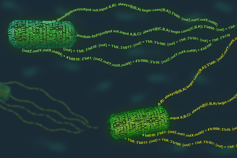
MIT researchers have developed synthetic biological circuits (from bacteria, for example, as shown here) that combine analog and digital computation as “living therapeutics” to treat major diseases and rare genetic disorders (credit: Synlogic)

MIT researchers have developed synthetic biological circuits that combine both analog (continuous) and digital (discrete) computation — allowing living cells to carry out complex processing operations, such as releasing a drug in response to low glucose levels.

The research is presented in an open-access paper published in the journal Nature Communications.


Background: analog vs. digital biological circuits

Like electronic circuits, living cells are capable of performing computations that are either continuous (analog) — like the way eyes adjust to gradual changes in the light levels — or digital, involving simple discrete on or off processes, such as a cell’s self-programmed death (apoptosis). Current synthetic biological systems, in contrast, have tended to focus on either analog or digital processing, limiting the range of uses.

Two basic logic circuits. An AND gate fires only if both inputs are “true” (for example, both inputs have a 1 volt signal, not zero). An OR gate fires if either (or both) of the inputs is true (for example, the top input has a 1 volt signal and the bottom input has zero. (credit: KurzweilAI)

Digital systems are based on a simple binary output, such as 0 or 1, so performing complex computational operations requires the use of a large number of parts (such as AND and OR logic gates) to make the decision, which is difficult to achieve in synthetic biological systems. (There are seven basic logic gates: AND, OR, XOR, NOT, NAND, NOR, and XNOR, as explained here.)

Using genes (instead of voltages), synthetic biologists design genetic circuits (arrangements of DNA components) that can perform new functions. For example, here’s a gene circuit that was constructed using Escherichia coli bacteria (source: Imperial College London/Nature Communications study):

Example of a biological AND gate. Two environment-responsive gene promoters (a region of DNA that initiates transcription — that is, copying a particular segment of DNA into RNA), P1 and P2, act as the inputs to drive the transcriptions of hrpR and hrpS genes, and respond to small molecules. Transcription of the output promoter gfp is turned on only when both proteins HrpR and HrpS are present. (credit: Baojun Wang et al./Nature Communications)


“Most of the work in synthetic biology has focused on the digital approach, because [digital systems] are much easier to program,” says Timothy Lu, an associate professor of electrical engineering and computer science and of biological engineering, and head of the Synthetic Biology Group at MIT’s Research Laboratory of Electronics.

The new synthetic circuits can measure the level of an analog input, such as a particular chemical relevant to a disease, and then make a binary decision — for example, turning on an output, such as a drug that treats the disease if the level is in the right range.

The new circuits are based on multiple elements. For example, a threshold module consists of a sensor that detects analog levels of a particular chemical, which controls the expression of the second digital component, a recombinase gene, which can then switch on or off a segment of DNA by converting it into a digital (on or off) output. (This conversion process is similar to electronic devices known as comparators, which take analog input signals and convert them into a digital output.)

If the concentration of the chemical reaches a certain level, the threshold module expresses the recombinase gene, causing it to flip the DNA segment (which contains a gene or gene-regulatory element, which then alters the expression of a desired output).

“So this is how we take an analog input, such as a concentration of a chemical, and convert it into a 0 or 1 signal,” Lu says. “And once that is done, and you have a piece of DNA that can be flipped upside down, then you can put together any of those pieces of DNA to perform digital computing,” he says.

Ternary logic for three-way glucose decisions

The team has also built an analog-to-digital converter circuit that implements ternary (three-valued) logic. The circuit, which is capable of producing two different outputs, will only switch on in response to either a high or low concentration range of an input.

In the future, the circuit could be used to detect glucose levels in the blood and respond in one of three ways depending on the concentration, he says. “If the glucose level was too high, you might want your cells to produce insulin, if the glucose was too low you might want them to make glucagon, and if it was in the middle you wouldn’t want them to do anything,” he says.

Similar analog-to-digital converter circuits could also be used to detect a variety of chemicals, simply by changing the sensor, Lu says.

Detecting inflammation and environmental conditions

The researchers are investigating the idea of using analog-to-digital converters to detect levels of inflammation in the gut caused by inflammatory bowel disease, for example, and releasing different amounts of a drug in response.

Immune cells used in cancer treatment could also be engineered to detect different environmental inputs, such as oxygen or tumor lysis (cell breakdown) levels, and vary the immune-call therapeutic activity in response.

Other research groups are also interested in using the devices for environmental applications, such as engineering cells that detect concentrations of water pollutants, Lu says.

The research team recently created a spinout company, called Synlogic, which is now attempting to use simple versions of the circuits to engineer probiotic bacteria that can treat diseases in the gut. The company hopes to begin clinical trials of these bacteria-based treatments within the next 12 months.


Abstract of Synthetic mixed-signal computation in living cells

Living cells implement complex computations on the continuous environmental signals that they encounter. These computations involve both analogue- and digital-like processing of signals to give rise to complex developmental programs, context-dependent behaviours and homeostatic activities. In contrast to natural biological systems, synthetic biological systems have largely focused on either digital or analogue computation separately. Here we integrate analogue and digital computation to implement complex hybrid synthetic genetic programs in living cells. We present a framework for building comparator gene circuits to digitize analogue inputs based on different thresholds. We then demonstrate that comparators can be predictably composed together to build band-pass filters, ternary logic systems and multi-level analogue-to-digital converters. In addition, we interface these analogue-to-digital circuits with other digital gene circuits to enable concentration-dependent logic. We expect that this hybrid computational paradigm will enable new industrial, diagnostic and therapeutic applications with engineered cells.

Scientists plan to build human genome from scratch

Efficiency trends in DNA sequencing (green) and synthesis of double-stranded DNA (dsDNA, blue) and single-stranded DNA (ssDNA, red) over the past ~35 years. The disruptive improvement in sequencing and ssDNA (oligonucleotides) synthesis technologies has improved from multiplex and miniaturization technologies in high-throughput DNA sequencing and oligo microarray technologies, respectively. (credit: Jef D. Boeke et al./Science)

Leading genomics experts have announced Genome Project-write (HGP-write), which aims to synthesize entire genomes of humans and other species from chemical components and get them to function in living cells.

As explained in Science, the goal of HGP-write is to reduce the costs of engineering large genomes, including a human genome, and to develop an ethical framework for genome-scale engineering and transformative medical applications.

Impacts expected on human health, energy, agriculture, chemicals, and bioremediation

HGP-write will build on the knowledge gained by The Human Genome Project (HGP-read), especially in genomic-based discovery, diagnostics, and therapeutics. But while the Human Genome Project “read” DNA to understand its code, HGP-write will use the cellular machinery provided by nature to “write” code, constructing vast DNA chains.

The goal is to launch HGP-write in 2016 with $100 million in committed support from public, private, philanthropic, industry, and academic sources globally. Autodesk has already contributed a leadership gift of $250,000 to seed the planning and launch of HGP-write.

According to the authors of the Science commentary, although “…sequencing, analyzing and editing DNA continues to advance at breakneck pace, the capability to construct DNA sequences in cells is mostly limited to a small number of short segments, restricting the ability to manipulate and understand biological systems.”

Exponential improvements in genome engineering

The new effort is expected to lead to a massive amount of information connecting the sequence of nucleotide bases in DNA with their physiological properties and functions, and it promises to have a significant impact on human health and other critical areas such as energy, agriculture, chemicals, and bioremediation, according to the organizers.

HGP-write will be implemented through a new, independent nonprofit organization, the Center of Excellence for Engineering Biology as an open, international, multi-disciplinary research project.*

“This grand challenge is more ambitious and more focused on understanding the practical applications than the original Human Genome Project,” said George Church, Ph.D., Robert Winthrop Professor of Genetics at Harvard Medical School. “Exponential improvements in genome engineering technologies and functional testing provide an opportunity to deepen the understanding of our genetic blueprint and use this knowledge to address many of the global problems facing humanity.”

Another goal is development of new genomics analysis, design, synthesis, assembly and testing technologies, with the goal of making them more affordable and widely available. “Writing DNA code is the future of science and medicine, but our technical capabilities remain limited,” said Andrew Hessel, Distinguished Researcher, Bio/Nano Research Group, Autodesk, who will head a multidisciplinary team exploring computer-aided design and manufacturing for biotechnology and nanotechnology R&D.

“HGP-write will require research and development on a grand scale, and this effort will help to push our current technical limits by several orders of magnitude,” he said.

The HGP-write project developed from a series of meetings held over the last several years, including a closed-door meeting held May 10 in Boston, which brought together a diverse group of 130 participants from many different countries, including biologists, ethicists, engineers, plus representatives from industry, law and government.

* The Genome Project-write (HGP-write) will be an open, international research project led by a multi-disciplinary group of scientific leaders who plan to oversee a reduction in the costs of engineering and testing large genomes, including a human genome, in cell lines more than 1,000-fold within ten years. 

To ensure public engagement and transparency, HGP-write will include mechanisms to encourage public discourse around the emerging HGP-write capabilities. The Woodrow Wilson Center for International Scholars will help to lead such efforts for HGP-write.

Principals are:

  • Jef Boeke, Ph.D., Director, Institute for Systems Genetics, Professor, Department of Biochemistry and Molecular Pharmacology, NYU Langone Medical Center. Dr. Boeke is a leader of the Synthetic Yeast Project (Sc2.0), which seeks to create living yeast cells with entirely redesigned chromosomes by 2017.
  • George Church, Ph.D., Robert Winthrop Professor of Genetics at Harvard Medical School, Core Faculty Member at the Wyss Institute for Biologically Inspired Engineering at Harvard University, Professor of Health Sciences and Technology at Harvard and the Massachusetts Institute of Technology (MIT), and Senior Associate Faculty member at the Broad Institute. Among the leaders of the original HGP-read, Dr. Church currently heads an effort to create a version of the bacteria E.coli with a redesigned genome.
  • Andrew Hessel, Distinguished Researcher, Bio/Nano Research Group, Autodesk. He spearheads a multidisciplinary team exploring computer-aided design and manufacturing for biotechnology and nanotechnology R&D.
  • Nancy J Kelley, J.D., M.P.P., President & CEO, Nancy J Kelley & Associates, formerly Founding Executive Director, New York Genome Center. She is lead executive of HGP-write and the related Center of Excellence for Engineering Biology.

Abstract of The Genome Project–Write

The Human Genome Project (“HGP-read”) nominally completed in 2004 aimed to sequence the human genome and improve technology, cost, and quality of DNA sequencing (12). It was biology’s first genome-scale project, and at the time was considered controversial by some. Now it is recognized as one of the great feats of exploration, one that has revolutionized science and medicine.

Although sequencing, analyzing, and editing DNA continue to advance at breakneck pace, the capability to construct DNA sequences in cells is mostly limited to a small number of short segments, restricting the ability to manipulate and understand biological systems. Further understanding of genetic blueprints could come from construction of large, gigabase (Gb)–sized animal and plant genomes, including the human genome, which would in turn drive development of tools and methods to facilitate large-scale synthesis and editing of genomes. To this end, we propose the Human Genome Project–Write (HGP-write).

Hacking life: how to program new functions for living bacteria and yeast

MIT biological engineers have devised a programming language that can be used to add new functions to E. coli bacteria (credit: Janet Iwasa)

MIT biological engineers have created a programming language for bacteria. It allows anyone to rapidly design complex, DNA-encoded circuits that add new functions to living cells — no genetic engineering knowledge required.

For example: design bacterial cells that can produce a cancer drug when they detect a tumor or create yeast cells that can halt their own fermentation process if too many toxic byproducts build up.

Repurposing a computer-chip programming language

The language is based on Verilog, a text-based language commonly used to program computer chips. To create a version of the language that would work for cells, the researchers designed computing elements such as logic gates and sensors that can be encoded in a bacterial cell’s DNA.

The sensors can detect different compounds, such as oxygen or glucose, and environmental conditions like light, temperature, and acidity. You can also add your own sensors, and you can compile a program for different organisms to get the right DNA sequence for each one.

You also specify the sensors, actuators, and a user constraints file (UCF), which defines the organism, gate technology, and valid operating conditions.

Cello (think of it as a compiler), a website, then uses this information to automatically design a DNA sequence encoding the desired circuit, using a set of algorithms that parse the Verilog text, create the circuit diagram, assign gates, balance constraints to build the DNA, and simulate performance, drawing on a library of Boolean logic gates.

Overview of Cello. Cello users write Verilog code and select or upload sensors and a UCF. On the basis of the Verilog design, a truth table is constructed, from which a circuit diagram is synthesized. Regulators are assigned from a library to each gate (each color is a different repressor). Combinatorial design is then used to concatenate parts into a linear DNA sequence. (credit: Alec A. K. Nielsen et al./Science)

In the current Verilog version, the genetic parts are optimized for E. coli, but the researchers are working on expanding the language for other strains of bacteria, including Bacteroides, commonly found in the human gut; Pseudomonas,which often lives in plant roots; and the yeast Saccharomyces cerevisiae.

Christopher Voigt, an MIT professor of biological engineering, and colleagues at Boston University and the National Institute of Standards and Technology have used this language, which they describe in the April 1 issue of Science.

No experience needed

Over the past 15 years, biologists and engineers have designed many genetic parts, such as sensors, memory switches, and biological clocks, that can be combined to modify existing cell functions and add new ones. However, designing each circuit is a laborious process that requires great expertise and often a lot of trial and error. “You have to have this really intimate knowledge of how those pieces are going to work and how they’re going to come together,” Voigt says.

Users of the new programming language, however, need no special knowledge of genetic engineering.

“You could be completely naive as to how any of it works. That’s what’s really different about this,” Voigt says. “You could be a student in high school and go onto the Web-based server and type out the program you want, and it spits back the DNA sequence.”

Using this language, the MIT researchers programmed 60 circuits with different functions, and 45 of them worked correctly the first time they were tested. Many of the circuits were designed to measure one or more environmental conditions, such as oxygen level or glucose concentration, and respond accordingly. Another circuit was designed to rank three different inputs and then respond based on the priority of each one.

One of the new circuits is the largest biological circuit ever built, containing seven logic gates and about 12,000 base pairs of DNA.

Another advantage of this technique is its speed. Until now, “it would take years to build these types of circuits. Now you just hit the button and immediately get a DNA sequence to test,” Voigt says.

His team plans to work on several different applications using this approach: bacteria that can be swallowed to aid in digestion of lactose; bacteria that can live on plant roots and produce insecticide if they sense the plant is under attack; and yeast that can be engineered to shut off when they are producing too many toxic byproducts in a fermentation reactor.


Abstract of Genetic circuit design automation

Computation can be performed in living cells by DNA-encoded circuits that process sensory information and control biological functions. Their construction is time-intensive, requiring manual part assembly and balancing of regulator expression. We describe a design environment, Cello, in which a user writes Verilog code that is automatically transformed into a DNA sequence. Algorithms build a circuit diagram, assign and connect gates, and simulate performance. Reliable circuit design requires the insulation of gates from genetic context, so that they function identically when used in different circuits. We used Cello to design 60 circuits for Escherichia coli (880,000 base pairs of DNA), for which each DNA sequence was built as predicted by the software with no additional tuning. Of these, 45 circuits performed correctly in every output state (up to 10 regulators and 55 parts), and across all circuits 92% of the output states functioned as predicted. Design automation simplifies the incorporation of genetic circuits into biotechnology projects that require decision-making, control, sensing, or spatial organization.

Craig Venter’s team designs, builds first minimal synthetic bacterial cell

A cluster of cells of a new synthetic species known as JCVI-syn3.0, showing spherical structures of varying sizes (scale bar, 200 nm). (credit: Clyde A. Hutchison III et al./Science)

Just 473 genes were needed to create life in a new synthesized species of bacteria created by synthetic biologists from the J. Craig Venter Institute (JCVI) and Synthetic Genomics, Inc.

Knowing the minimum number of genes to create life would answer a fundamental question in biology.

This “minimal synthetic cell,” JCVI-syn3.0, was reported in an open-access paper published last week in the  journal Science. By comparison, the first synthetic cell developed by the scientists, M. mycoides JCVI-syn1.0, has 1.08 million base pairs and 901 genes.*

The new cell contains 531,560 base pairs (the “alphabet” or sequence that makes up the DNA code) and 473 genes — the smallest number of genes of any organism that can be grown in a laboratory, according to the team.


JCVI | CVI-syn3.0 — Minimal Cell

“All of the…studies over the past 20 years have underestimated the number of essential genes by focusing only on the known world. This is an important observation that we are carrying forward into the study of the human genome,” said senior author and group leader J. Craig Venter, PhD.

For 50 years, researchers have studied essential and non-essential genes in bacteria to help biologists understand the core functions needed for life. In the newer field of synthetic biology, this same information will be able to help scientists design DNA sequences for new synthetic organisms — allowing them to build frameworks for industrial applications of synthetic organisms.

The cycle for genome design, building by means of synthesis and cloning in yeast, and testing for viability by means of genome transplantation. After each cycle, gene essentiality is reevaluated by global transposon mutagenesis. (credit: Clyde A. Hutchison III et al./Science)

Mystery genes

During construction** of JCVI-syn3.0, the team discovered that 149 of the genes actually had unknown functions, but were essential for robust growth. Those functions remain an area of continued work for the researchers. Other genes in the minimal synthetic cell were related to reading and expressing the DNA code, structure, and function of the outer cell membrane, and cell metabolism, or to preserving DNA integrity.

However, the team expects to decode the 149 genes in the future. “Our goal is to have a cell for which the precise biological function of every gene is known,” said Clyde Hutchison, PhD, first author and distinguished professor at JCVI.

Another major outcome of the minimal cell program will be new tools and semi-automated processes for synthesizing the DNA sequence needed for whole organisms, according to Daniel Gibson, PhD, an associate professor at JCVI.

“This paper signifies a major step toward our ability to design and build synthetic organisms from the bottom up with predictable outcomes,” he said. “The tools and knowledge gained from this work will be essential to producing next-generation production platforms for a wide range of disciplines.”

This work was funded by SGI, the JCVI endowment, and the Defense Advanced Research Projects Agency’s Living Foundries program.

* The research at JCVI leading to this report began in 1995 with DNA sequencing of the first free-living organism, Haemophilus influenza, followed by the DNA sequencing of Mycoplasma genitalium. A comparison of these two genomes revealed a common set of 256 genes that the team thought could be a minimal set of genes needed for viability.

In 1999, Hutchison led a team who published a paper describing techniques to identify the non-essential genes in M. genitalium.

The creation of the first synthetic cell (JCVI-syn1.0) in 2010 established a workflow for building and testing designs for the DNA of a whole organism. This included design of a minimal cell from the bottom up, starting with the DNA sequence.

** To create JCVI-syn3.0, the team used an approach of whole genome design (design of the DNA needed for a whole organism) and chemical synthesis followed by genome transplantation to test if the cell was viable. Their first attempt to minimize the genome began with a simple approach using information in the biochemical literature and some limited mutations of DNA, but this did not result in a viable genome. After improving methods, the team discovered a set of “quasi-essential” genes that are necessary for robust growth and that explained the failure of their first attempt.

The team built the genome in eight segments at a time so that each could be tested separately before combining them to generate a minimal genome. The team also explored the order of the genes and how that affects cell growth and viability. They found gene content was more critical than gene order. They went through three cycles of designing, building, and testing ensuring that the “quasi-essential” genes remained, which in the end resulted in a viable, self-replicating minimal synthetic cell that contained just 473 genes.


Abstract of Design and synthesis of a minimal bacterial genome

We used whole-genome design and complete chemical synthesis to minimize the 1079–kilobase pair synthetic genome of Mycoplasma mycoides JCVI-syn1.0. An initial design, based on collective knowledge of molecular biology combined with limited transposon mutagenesis data, failed to produce a viable cell. Improved transposon mutagenesis methods revealed a class of quasi-essential genes that are needed for robust growth, explaining the failure of our initial design. Three cycles of design, synthesis, and testing, with retention of quasi-essential genes, produced JCVI-syn3.0 (531 kilobase pairs, 473 genes), which has a genome smaller than that of any autonomously replicating cell found in nature. JCVI-syn3.0 retains almost all genes involved in the synthesis and processing of macromolecules. Unexpectedly, it also contains 149 genes with unknown biological functions. JCVI-syn3.0 is a versatile platform for investigating the core functions of life and for exploring whole-genome design.

3D-printing a new lifelike liver tissue for drug screening

Images of the 3D-printed parts of the biomimetic liver tissue: liver cells derived from human induced pluripotent stem cells (left), endothelial and mesenchymal supporing cells (center), and the resulting organized combination of multiple cell types (right). (credit: Chen Laboratory, UC San Diego)

University of California, San Diego researchers have 3D-printed a tissue that closely mimics the human liver’s sophisticated structure and function. The new model could be used for patient-specific drug screening and disease modeling and could help pharmaceutical companies save time and money when developing new drugs, according to the researchers.

The liver plays a critical role in how the body metabolizes drugs and produces key proteins, so liver models are increasingly being developed in the lab as platforms for drug screening. However, so far, the models lack both the complex micro-architecture and diverse cell makeup of a real liver. For example, the liver receives a dual blood supply with different pressures and chemical constituents.

So the team employed a novel bioprinting technology that can rapidly produce complex 3D microstructures that mimic the sophisticated features found in biological tissues.

3D bioprinting of hydrogel-based hepatic (liver) construct. (A) Schematic diagram of a two-step 3D-bioprinting approach in which hiPSC-HPCs (human induced pluripotent stem cell-derived hepatic progenitor cells) were patterned by the first digital mask, followed by the patterning of supporting cells using a second digital mask. (B) Grayscale digital masks corresponding to polymerizing lobule structure (Left) and vascular structure (Right) designed for two-step bioprinting. The white patterns represent the light-reflecting patterns for photo-polymerization. (C) Images (5x) taken under fluorescent and bright field channels showing patterns of hiPSC-HPCs  (green) and supporting cells (red). (credit: Xuanyi Ma et al./PNAS)

The liver tissue was printed in two steps.

  • The team printed a honeycomb pattern of 900-micrometer-sized hexagons, each containing liver cells derived from human induced pluripotent stem cells. An advantage of human induced pluripotent stem cells is that they are patient-specific, which makes them ideal materials for building patient-specific drug screening platforms. And since these cells are derived from a patient’s own skin cells, researchers don’t need to extract any cells from the liver to build liver tissue.
  • Then, endothelial and mesenchymal supporting cells were printed in the spaces between the stem-cell-containing hexagons.

The entire structure — a 3 × 3 millimeter square, 200 micrometers thick — takes just seconds to print. The researchers say this is a vast improvement over other methods to print liver models, which typically take hours. Their printed model was able to maintain essential functions over a longer time period than other liver models. It also expressed a relatively higher level of a key enzyme that’s considered to be involved in metabolizing many of the drugs administered to patients.

“It typically takes about 12 years and $1.8 billion to produce one FDA-approved drug,” said Shaochen Chen, NanoEngineering professor at the UC San Diego Jacobs School of Engineering. “That’s because over 90 percent of drugs don’t pass animal tests or human clinical trials. We’ve made a tool that pharmaceutical companies could use to do pilot studies on their new drugs, and they won’t have to wait until animal or human trials to test a drug’s safety and efficacy on patients. This would let them focus on the most promising drug candidates earlier on in the process.”

The work was published the week of Feb. 8 in the online early edition of Proceedings of the National Academy of Sciences.


Abstract of Deterministically patterned biomimetic human iPSC-derived hepatic model via rapid 3D bioprinting

The functional maturation and preservation of hepatic cells derived from human induced pluripotent stem cells (hiPSCs) are essential to personalized in vitro drug screening and disease study. Major liver functions are tightly linked to the 3D assembly of hepatocytes, with the supporting cell types from both endodermal and mesodermal origins in a hexagonal lobule unit. Although there are many reports on functional 2D cell differentiation, few studies have demonstrated the in vitro maturation of hiPSC-derived hepatic progenitor cells (hiPSC-HPCs) in a 3D environment that depicts the physiologically relevant cell combination and microarchitecture. The application of rapid, digital 3D bioprinting to tissue engineering has allowed 3D patterning of multiple cell types in a predefined biomimetic manner. Here we present a 3D hydrogel-based triculture model that embeds hiPSC-HPCs with human umbilical vein endothelial cells and adipose-derived stem cells in a microscale hexagonal architecture. In comparison with 2D monolayer culture and a 3D HPC-only model, our 3D triculture model shows both phenotypic and functional enhancements in the hiPSC-HPCs over weeks of in vitro culture. Specifically, we find improved morphological organization, higher liver-specific gene expression levels, increased metabolic product secretion, and enhanced cytochrome P450 induction. The application of bioprinting technology in tissue engineering enables the development of a 3D biomimetic liver model that recapitulates the native liver module architecture and could be used for various applications such as early drug screening and disease modeling.

Cell-free protein synthesis device is potential lifesaver

This section of a serpentine channel reactor shows the parallel reactor and feeder channels separated by a nanoporous membrane. Left: a single nanopore viewed from the side; right: a diagram of metabolite exchange across the membrane. (credit: ORNL)

Oak Ridge National Laboratory scientists have developed a device that uses microfabricated bioreactors to produce therapeutic proteins for medicines and biopharmaceuticals. These miniature factories are cell-free, eliminating the need to maintain a living system, which radically simplifies the process and lowers cost, and makes the device easily adaptable for use in isolated locations and at disaster sites.

On-demand, point-of-care therapeutic protein synthesis requires that a dose of protein be produced and purified quickly. “With this approach, we can produce more protein faster, making our technology ideal for point-of-care use,” said team co-leader Scott Retterer of the lab’s Biosciences Division. It can produce proteins “when and where you need them, bypassing the challenge of keeping the proteins cold during shipment and storage.”

The key to the cell-free reactions in the new bioreactor is a permeable nanoporous membrane and serpentine (snake-like) design, made using a combination of electron-beam lithography and advanced material-deposition processes.

The long serpentine channels allow for exchange of materials between parallel reactor and feeder channels. With this approach, the team can control the exchange of metabolites, energy, and species that inhibit production of the desired protein. The design also extends reaction times and improves yields.

“We show that the microscale bioreactor design produces higher protein yields than conventional tube-based batch formats and that product yields can be dramatically improved by facilitating small molecule exchange with the dual-channel bioreactor,” the authors wrote in their paper, published in the journal Small.

The researchers also note that on-demand biologic synthesis would aid production of drugs that are costly to mass-produce, including orphan drugs and personalized medicines.


Abstract of Toward Microfluidic Reactors for Cell-Free Protein Synthesis at the Point-of-Care

Cell-free protein synthesis (CFPS) is a powerful technology that allows for optimization of protein production without maintenance of a living system. Integrated within micro and nanofluidic architectures, CFPS can be optimized for point-of-care use. Here, the development of a microfluidic bioreactor designed to facilitate the production of a single-dose of a therapeutic protein, in a small footprint device at the point-of-care, is described. This new design builds on the use of a long, serpentine channel bioreactor and is enhanced by integrating a nanofabricated membrane to allow exchange of materials between parallel “reactor” and “feeder” channels. This engineered membrane facilitates the exchange of metabolites, energy, and inhibitory species, and can be altered by plasma-enhanced chemical vapor deposition and atomic layer deposition to tune the exchange rate of small molecules. This allows for extended reaction times and improved yields. Further, the reaction product and higher molecular weight components of the transcription/translation machinery in the reactor channel can be retained. It has been shown that the microscale bioreactor design produces higher protein yields than conventional tube-based batch formats, and that product yields can be dramatically improved by facilitating small molecule exchange within the dual-channel bioreactor.