Omnidirectional wireless charging up to half a meter away from a power source

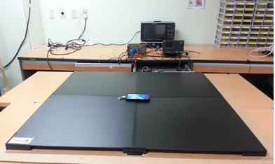
Omnidirectional wireless-charging system can charge multiple numbers of mobile devices simultaneously in a one-cubic-meter range. Above: charging transmitter; below: a Samsung Galaxy Note with embedded receiver. (credit: KAIST)

A group of researchers at KAIST in Korea has developed a wireless-power transfer (WPT) technology that allows mobile devices in the “Wi-Power” zone (within 0.5 meters from the power source) to be charged at any location and in any direction and orientation, tether-free.

The WPT system is capable of charging 30 smartphones with a power capacity of one watt each or 5 laptops with 2.4 watts.

The research team used its Dipole Coil Resonance System (DCRS) to induce magnetic fields, composed of two (transmitting and receiving) magnetic dipole coils, placed in parallel. Each coil has a ferrite core and is connected with a resonant capacitor.

Current wireless-power technologies require close contact with a charging pad and are limited to a fixed position.

The research was published in the June 2015 on-line issue of IEEE Transactions on Power Electronics.


KAIST | KAIST Omnidirectional Wireless Smartphone Charger at 1m


Abstract of Six Degrees of Freedom Mobile Inductive Power Transfer by Crossed Dipole Tx and Rx Coils

Crossed dipole coils for the wide-range 3-D omnidirectional inductive power transfer (IPT) are proposed. Free positioning of a plane receiving (Rx) coil is obtained for an arbitrary direction within 1m from a plane transmission (Tx) coil. Both the Tx and Rx coils consist of crossed dipole coils with an orthogonal phase difference; hence, a rotating magnetic field is generated from the Tx, which enables the Rx to receive power vertically or horizontally. Thus, the 3-D omnidirectional IPT is first realized for both the plate type Tx and Rx coils, which is crucial for practical applications where volumetric coil structure is highly prohibited. This optimized configuration of coils has been obtained through a general classification of power transfer and searching for mathematical constraints on multi-D omnidirectional IPT. Conventional loop coils are thoroughly analyzed and verified to be inadequate for the plate-type omnidirectional IPT in this paper. Simulation-based design of the proposed crossed dipole coils for a uniform magnetic field distribution is provided, and the 3-D omnidirectional IPT is experimentally verified by prototype Rx coils for a wireless power zone of 1 m3 with a prototype Tx coil of 1 m2 at an operating frequency of 280 kHz, meeting the Power Matters Alliance (PMA). The maximum overall efficiency was 33.6% when the input power was 100 W.

AI algorithm learns to ‘see’ features in galaxy images

Hubble Space Telescope image of the cluster of galaxies MACS0416.1-2403, one of the Hubble “Frontier Fields” images. Bright yellow “elliptical” galaxies can be seen, surrounded by numerous blue spiral and amorphous (star-forming) galaxies. This image forms the test data that the machine learning algorithm is applied to, having not previously “seen” the image. (credit: NASA/ESA/J. Geach/A. Hocking)

A team of astronomers and computer scientists at the University of Hertfordshire have taught a machine to “see” astronomical images, using data from the Hubble Space Telescope Frontier Fields set of images of distant clusters of galaxies that contain several different types of galaxies.

The technique, which uses a form of AI called unsupervised machine learning, allows galaxies to be automatically classified at high speed, something previously done by thousands of human volunteers in projects like Galaxy Zoo.

Image highlighting parts of the MACS0416.1-2403 cluster image that the algorithm has identified as “star-forming” galaxies (credit: NASA/ESA/J. Geach/A. Hocking)

“We have not told the machine what to look for in the images, but instead taught it how to ‘see,’” said graduate student Alex Hocking.

“Our aim is to deploy this tool on the next generation of giant imaging surveys where no human, or even group of humans, could closely inspect every piece of data. But this algorithm has a huge number of applications far beyond astronomy, and investigating these applications will be our next step,” said University of Hertfordshire Royal Society University Research Fellow James Geach, PhD.

The scientists are now looking for collaborators to make use of the technique in applications like medicine, where it could for example help doctors to spot tumors, and in security, to find suspicious items in airport scans.

Your entire viral infection history from a single drop of blood

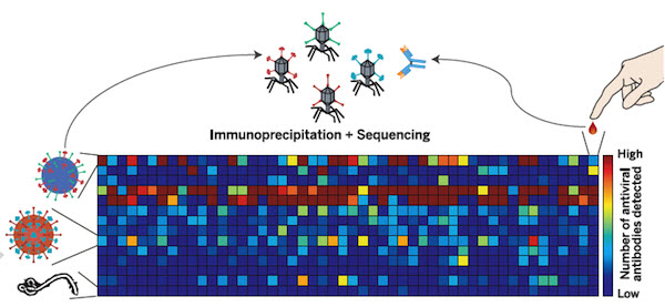
Systematic viral epitope scanning (VirScan). This method allows comprehensive analysis of antiviral antibodies in human sera. VirScan combines DNA microarray synthesis and bacteriophage display to create a uniform, synthetic representation of peptide epitopes comprising the human virome. Immunoprecipitation and high-throughput DNA sequencing reveal the peptides recognized by antibodies in the sample. The color of each cell in the heatmap depicts the relative number of antigenic epitopes detected for a virus (rows) in each sample (columns). (credit: George J. Xu et al./Science).

New technology called called VirScan developed by Howard Hughes Medical Institute (HHMI) researchers makes it possible to test for current and past infections with any known human virus by analyzing a single drop of a person’s blood.

With VirScan, scientists can run a single test to determine which viruses have infected an individual, rather than limiting their analysis to particular viruses. That unbiased approach could uncover unexpected factors affecting individual patients’ health, and also expands opportunities to analyze and compare viral infections in large populations.

The comprehensive analysis can be performed for about $25 per blood sample, but the test is currently being used only as a research tool and is not yet commercially available.

Stephen J. Elledge, an HHMI investigator at Brigham and Women’s Hospital, led the development of VirScan. He and his colleagues have already used VirScan to screen the blood of 569 people in the United States, South Africa, Thailand, and Peru. The scientists described the new technology and reported their findings in the June 5, 2015, issue of the journal Science.

Virus antibodies: clues that last decades
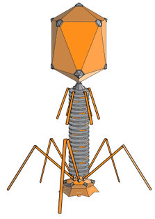
Bacteriophage (credit: Wikimedia Commons)

VirScan works by screening the blood for antibodies against any of the 206 species of viruses known to infect humans*. The immune system ramps up production of pathogen-specific antibodies when it encounters a virus for the first time, and it can continue to produce those antibodies for years or decades after it clears an infection.

That means VirScan not only identifies viral infections that the immune system is actively fighting, but also provides a history of an individual’s past infections.

To develop the new test, Elledge and his colleagues synthesized more than 93,000 short pieces of DNA encoding different segments of viral proteins. They introduced those pieces of DNA into bacteria-infecting viruses called bacteriophage.

Each bacteriophage manufactured one of the protein segments — known as a peptide — and displayed the peptide on its surface. As a group, the bacteriophage displayed all of the protein sequences found in the more than 1,000 known strains of human viruses.

To test the method, the team used it to analyze blood samples from patients known to be infected with particular viruses, including HIV and hepatitis C. “It turns out that it works really well,” Elledge says. “We were in the sensitivity range of 95 to 100 percent for those, and the specificity was good—we  didn’t falsely identify people who were negative. That gave us confidence that we could detect other viruses, and when we did see them we would know they were real.”


Harvard Medical School | Viral History in a Drop of Blood

International study

Elledge and his colleagues used VirScan to analyze the antibodies in 569 people from four countries, examining about 100 million potential antibody/epitope interactions. They found that on average, each person had antibodies to ten different species of viruses. As expected, antibodies against certain viruses were common among adults but not in children, suggesting that children had not yet been exposed to those viruses. Individuals residing South Africa, Peru, and Thailand, tended to have antibodies against more viruses than people in the United States. The researchers also found that people infected with HIV had antibodies against many more viruses than did people without HIV.

Elledge says the team was surprised to find that antibody responses against specific viruses were surprisingly similar between individuals, with different people’s antibodies recognizing identical  amino acids in the viral peptides. “In this paper alone we identified more antibody/peptide interactions to viral proteins than had been identified in the previous history of all viral exploration,” he says. The surprising reproducibility of those interactions allowed the team to refine their analysis and improve the  sensitivity of VirScan, and Elledge says the method will continue to improve as his team analyzes more samples. Their findings on viral epitopes may also have important implications for vaccine design.

Elledge says the approach his team has developed is not limited to antiviral antibodies. His own lab is also using it to look for antibodies that attack a body’s own tissue in certain autoimmune diseases that are associated with cancer. A similar approach could also be used to screen for antibodies against other types of pathogens.

* Antibodies in the blood find their viral targets by recognizing unique features known as epitopes that are embedded in proteins on the virus surface. To perform the VirScan analysis, all of the peptide-displaying bacteriophage are allowed to mingle with a blood sample. Antiviral antibodies in the blood find and bind to their target epitopes within the displayed peptides. The scientists then retrieve the antibodies and wash away everything except for the few bacteriophage that cling to them. By sequencing the DNA of those bacteriophage, they can identify which viral protein pieces were grabbed onto by antibodies in the blood sample. That tells the scientists which viruses a person’s immune system has previously encountered, either through infection or through vaccination. Elledge estimates it would take about 2-3 days to process 100 samples, assuming sequencing is working optimally. He is optimistic the speed of the assay will increase with further development.


Abstract of Comprehensive serological profiling of human populations using a synthetic human virome

The human virome plays important roles in health and immunity. However, current methods for detecting viral infections and antiviral responses have limited throughput and coverage. Here, we present VirScan, a high-throughput method to comprehensively analyze antiviral antibodies using immunoprecipitation and massively parallel DNA sequencing of a bacteriophage library displaying proteome-wide peptides from all human viruses. We assayed over 108 antibody-peptide interactions in 569 humans across four continents, nearly doubling the number of previously established viral epitopes. We detected antibodies to an average of 10 viral species per person and 84 species in at least two individuals. Although rates of specific virus exposure were heterogeneous across populations, antibody responses targeted strongly conserved “public epitopes” for each virus, suggesting that they may elicit highly similar antibodies. VirScan is a powerful approach for studying interactions between the virome and the immune system.
Creating DNA-based nanostructures without water

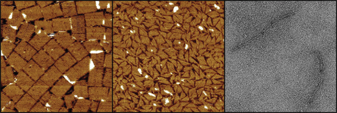
Three different DNA nanostructures assembled at room temperature in water-free glycholine (left) and in 75 percent glycholine-water mixture (center and right). The structures are (from left to right) a tall rectangle two-dimensional DNA origami, a triangle made of single-stranded tails, and a six-helix bundle three-dimensional DNA origami (credit: Isaac Gállego).

Researchers at the Georgia Institute of Technology have discovered an new process for assembling DNA nanostructures in a water-free solvent, which may allow for fabricating more complex nanoscale structures — especially, nanoelectronic chips based on DNA.

Scientists have been using DNA to construct sophisticated new structures from nanoparticles (such as a recent development at Brookhaven National Labs reported by KurzweilAI May 26), but the use of DNA has required a water-based environment. That’s because DNA naturally functions inside the watery environment of living cells. However, the use of water limited the types of structures that are possible.

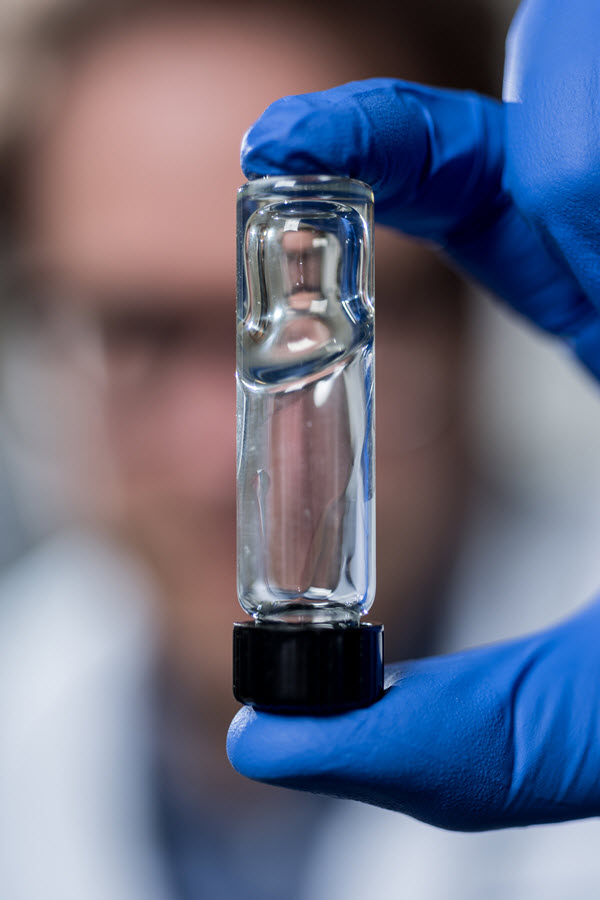
The viscosity of a new solvent used for assembling DNA nanostructures (credit: Rob Felt)

In addition, the Georgia Tech researchers discovered that, paradoxically, adding a small amount of water to their water-free solvent during the assembly process (and removing it later) increases the assembly rate. It could also allow for even more complex structures, by reducing the problem of DNA becoming trapped in unintended structures by aggregation (clumping).

The new solvent they used is known as glycholine, a mixture of glycerol (used for sweetening and preserving food) and choline chloride, but the researchers are exploring other materials.

The solvent system could improve the combined use of metallic nanoparticles and DNA based materials at room temperature. The solvent’s low volatility could also allow for storage of assembled DNA structures without the concern that a water-based medium would dry out.

The research on water-free solvents grew out of Georgia Tech researchers’ studies in the origins of life. They wondered if the molecules necessary for life, such as the ancestor of DNA, could have developed in a water-free solution. In some cases, they found, the chemistry necessary to make the molecules of life would be much easier without water being present.

Sponsored by the National Science Foundation and NASA, the research will be published as the cover story in Volume 54, Issue 23 of the journal Angewandte Chemie International Edition.

* The assembly rate of DNA nanostructures can be very slow, and depends strongly on temperature. Raising the temperature increases this rate, but temperatures that are too high can cause the DNA structures to fall apart. The solvent system developed at Georgia Tech adds a new level of control over DNA assembly. DNA structures assemble at lower temperatures in this solvent, and adding water can adjust the solvent’s viscosity (resistance to flow), which allows for faster assembly compared to the water-free version of the solvent.


Abstract of Folding and Imaging of DNA Nanostructures in Anhydrous and Hydrated Deep-Eutectic Solvents

There is great interest in DNA nanotechnology, but its use has been limited to aqueous or substantially hydrated media. The first assembly of a DNA nanostructure in a water-free solvent, namely a low-volatility biocompatible deep-eutectic solvent composed of a 4:1 mixture of glycerol and choline chloride (glycholine), is now described. Glycholine allows for the folding of a two-dimensional DNA origami at 20 °C in six days, whereas in hydrated glycholine, folding is accelerated (≤3 h). Moreover, a three-dimensional DNA origami and a DNA tail system can be folded in hydrated glycholine under isothermal conditions. Glycholine apparently reduces the kinetic traps encountered during folding in aqueous solvent. Furthermore, folded structures can be transferred between aqueous solvent and glycholine. It is anticipated that glycholine and similar solvents will allow for the creation of functional DNA structures of greater complexity by providing a milieu with tunable properties that can be optimized for a range of applications and nanostructures.

South Korean Team Kaist wins DARPA Robotics Challenge

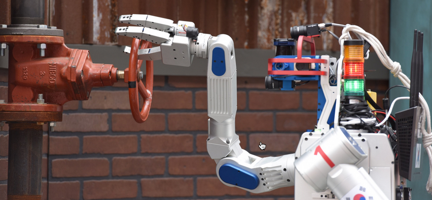
DRC-Hubo robot turns valve 360 degrees in DARPA Robotics Challenge Final (credit: DARPA)

First place in the DARPA Robotics Challenge Finals this past weekend in Pomona, California went to Team Kaist of South Korea for its DRC-Hubo robot, winning $2 million in prize money.

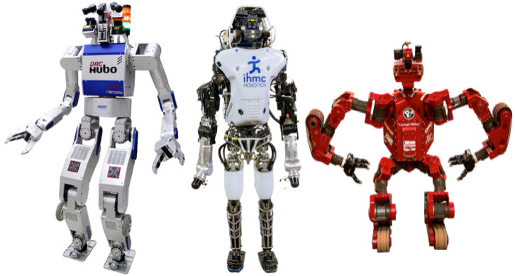
Team IHMC Robotics of Pensacola, Fla., with its Running Man (Atlas) robot came in at second place ($1 million prize), followed by Tartan Rescue of Pittsburgh with its CHIMP robot ($500,000 prize).

DRC-Hubo, Running Man, and CHIMP (credit: DARPA)

The DARPA Robotics Challenge, with three increasingly demanding competitions over two years, was launched in response to a humanitarian need that became glaringly clear during the nuclear disaster at Fukushima, Japan, in 2011, DARPA said.

The goal was to “accelerate progress in robotics and hasten the day when robots have sufficient dexterity and robustness to enter areas too dangerous for humans and mitigate the impacts of natural or man-made disasters.”

The difficult course of eight tasks simulated Fukushima-like conditions, such as driving alone, walking through rubble, tripping circuit breakers, turning valves, and climbing stairs.

Representing some of the most advanced robotics research and development organizations in the world, a dozen teams from the United States and another eleven from Japan, Germany, Italy, Republic of Korea and Hong Kong competed.

DARPA | DARPA Robotics Challenge 2015 Proving the Possible


DARPA | A Celebration of Risk (a.k.a., Robots Take a Spill)

More DARPA Robotics Challenge videos