Super-resolution electron microscopy of soft materials like biomaterials

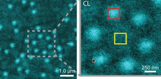
CLAIRE image of Al nanostructures with an inset that shows a cluster of six Al nanostructures (credit: Lawrence Berkeley National Laboratory)

Soft matter encompasses a broad swath of materials, including liquids, polymers, gels, foam and — most importantly — biomolecules. At the heart of soft materials, governing their overall properties and capabilities, are the interactions of nano-sized components.

Observing the dynamics behind these interactions is critical to understanding key biological processes, such as protein crystallization and metabolism, and could help accelerate the development of important new technologies, such as artificial photosynthesis or high-efficiency photovoltaic cells.

Observing these dynamics at sufficient resolution has been a major challenge, but this challenge is now being met with a new non-invasive nanoscale imaging technique that goes by the acronym of CLAIRE.

CLAIRE stands for “cathodoluminescence activated imaging by resonant energy transfer.” Invented by researchers with the U.S. Department of Energy (DOE)’s Lawrence Berkeley National Laboratory (Berkeley Lab) and the University of California (UC) Berkeley, CLAIRE extends the extremely high resolution of electron microscopy to the dynamic imaging of soft matter.

“Traditional electron microscopy damages soft materials and has therefore mainly been used to provide topographical or compositional information about robust inorganic solids or fixed sections of biological specimens,” says chemist Naomi Ginsberg, who leads CLAIRE’s development and holds appointments with Berkeley Lab’s Physical Biosciences Division and its Materials Sciences Division, as well as UC Berkeley’s departments of chemistry and physics.

“CLAIRE allows us to convert electron microscopy into a new non-invasive imaging modality for studying soft materials and providing spectrally specific information about them on the nanoscale.”

Ginsberg is also a member of the Kavli Energy NanoScience Institute (Kavli-ENSI) at Berkeley. She and her research group recently demonstrated CLAIRE’s imaging capabilities by applying the technique to aluminum nanostructures and polymer films that could not have been directly imaged with electron microscopy.

“What microscopic defects in molecular solids give rise to their functional optical and electronic properties? By what potentially controllable process do such solids form from their individual microscopic components, initially in the solution phase? The answers require observing the dynamics of electronic excitations or of molecules themselves as they explore spatially heterogeneous landscapes in condensed phase systems,” Ginsberg says.

“In our demonstration, we obtained optical images of aluminum nanostructures with 46 nanometer resolution, then validated the non-invasiveness of CLAIRE by imaging a conjugated polymer film. The high resolution, speed and non-invasiveness we demonstrated with CLAIRE positions us to transform our current understanding of key biomolecular interactions.”

How to avoid destroying soft matter with electron beams

CLAIRE works by essentially combining the best attributes of optical and scanning electron microscopy into a single imaging platform.

Scanning electron microscopes use beams of electrons rather than light for illumination and magnification. With much shorter wavelengths than photons of visible light, electron beams can be used to observe objects hundreds of times smaller than those that can be resolved with an optical microscope. However, these electron beams destroy most forms of soft matter and are incapable of spectrally specific molecular excitation.

Ginsberg and her colleagues get around these problems by employing a process called “cathodoluminescence,” in which an ultrathin scintillating film, about 20 nanometers thick, composed of cerium-doped yttrium aluminum perovskite, is inserted between the electron beam and the sample.

When the scintillating film is excited by a low-energy electron beam (about 1 KeV), it emits energy that is transferred to the sample, causing the sample to radiate. This luminescence is recorded and correlated to the electron beam position to form an image that is not restricted by the optical diffraction limit (which limits optical microscopy).

The CLAIRE imaging demonstration was carried out at the Molecular Foundry, a DOE Office of Science User Facility.

Observing biomolecular interactions, solar cells, and LEDs

While there is still more work to do to make CLAIRE widely accessible, Ginsberg and her group are moving forward with further refinements for several specific applications.

“We’re interested in non-invasively imaging soft functional materials like the active layers in solar cells and light-emitting devices,” she says. “It is especially true in organics and organic/inorganic hybrids that the morphology of these materials is complex and requires nanoscale resolution to correlate morphological features to functions.”

Ginsberg and her group are also working on the creation of liquid cells for observing biomolecular interactions under physiological conditions. Since electron microscopes can only operate in a high vacuum, as molecules in the air disrupt the electron beam, and since liquids evaporate in high vacuum, aqueous samples must either be freeze-dried or hermetically sealed in special cells.

“We need liquid cells for CLAIRE to study the dynamic organization of light-harvesting proteins in photosynthetic membranes,” Ginsberg says. “We should also be able to perform other studies in membrane biophysics to see how molecules diffuse in complex environments, and we’d like to be able to study molecular recognition at the single molecule level.”

In addition, Ginsberg and her group will be using CLAIRE to study the dynamics of nanoscale systems for soft materials in general. “We would love to be able to observe crystallization processes or to watch a material made of nanoscale components anneal or undergo a phase transition,” she says. “We would also love to be able to watch the electric double layer at a charged surface as it evolves, as this phenomenon is crucial to battery science.”

A paper describing the most recent work on CLAIRE has been published in the journal Nano Letters. This research was primarily supported by the DOE Office of Science and by the National Science Foundation.


Abstract of Cathodoluminescence-Activated Nanoimaging: Noninvasive Near-Field Optical Microscopy in an Electron Microscope

We demonstrate a new nanoimaging platform in which optical excitations generated by a low-energy electron beam in an ultrathin scintillator are used as a noninvasive, near-field optical scanning probe of an underlying sample. We obtain optical images of Al nanostructures with 46 nm resolution and validate the noninvasiveness of this approach by imaging a conjugated polymer film otherwise incompatible with electron microscopy due to electron-induced damage. The high resolution, speed, and noninvasiveness of this “cathodoluminescence-activated” platform also show promise for super-resolution bioimaging.

3-D printing tough biogel structures for tissue engineering or soft robots

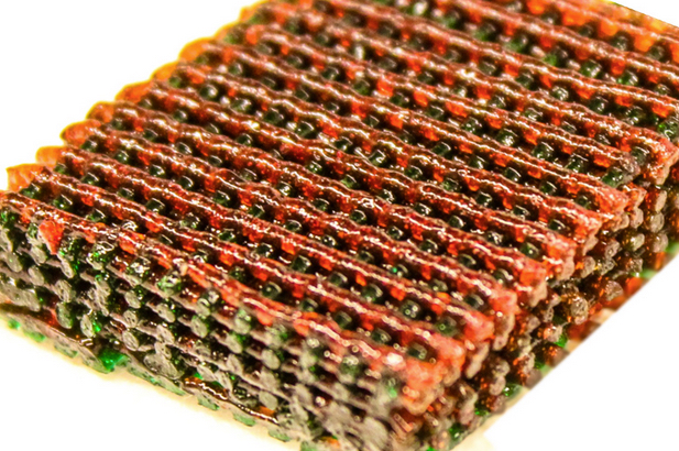
Lasagna? No, an open lattice of 3-D printed material, with materials having different characteristics of strength and flexibility indicated by different colors (credit: the researchers)

Researchers at three universities have developed a new way of making tough — but soft and wet — biocompatible hydrogel materials into complex and intricately patterned shapes. The process might lead to scaffolds for repair or replacement of load-bearing tissues, such as cartilage. It could also allow for tough but flexible actuators for future robots, the researchers say.

The new process is described in a paper in the journal Advanced Materials, co-authored by MIT associate professor of mechanical engineering Xuanhe Zhao and colleagues at MIT, Duke University, and Columbia University.

Zhao says the process can produce complex hydrogel structures that are “extremely tough and robust,” but still allow for encapsulating cells in the structures. That could make it possible to 3D-print complex biostructures.

Biocompatible structures

Hydrogels are defined by water molecules encased in rubbery polymer networks that provide shape and structure. They are similar to natural tissues such as cartilage, which is used by the body as a natural shock absorber.

While synthetic hydrogels are commonly weak or brittle, a number of them that are tough and stretchable have been developed over the last decade. However, making tough hydrogels has usually involved “harsh chemical environments” that would kill living cells encapsulated in them, Zhao says.

The new hydrogel materials are generated by combining polyethylene glycol (PEG) and sodium alginate, which synergize to form a hydrogel tougher than natural cartilage. The materials are benign enough to synthesize together with living cells — such as stem cells — which could then allow high viability of the cells, says Zhao, who holds a joint appointment in MIT’s Department of Civil and Environmental Engineering.

3-D printing strong, flexible biomaterials

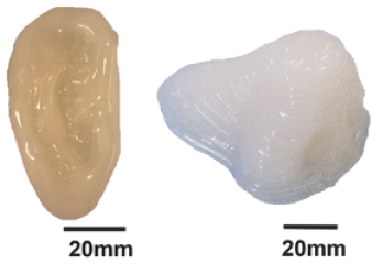
3-D printed tough, biocompatible PEG–alginate–nanoclay hydrogels in ear and nose shapes (credit: Sungmin Hong et al./ Advanced Materials)

Previous work was not able to produce complex 3-D structures with tough hydrogels, Zhao says. The new biocompatible tough hydrogel can be printed into diverse 3-D structures such as a hollow cube, hemisphere, pyramid, twisted bundle, multilayer mesh, or physiologically relevant shapes, such as a human nose or ear.

The new method uses a commercially available 3D-printing mechanism, Zhao explains. “The innovation is really about the material — a new ink for 3-D printing of biocompatible tough hydrogel,” he says, specifically, a composite of two different biopolymers.

“Each [material] individually is very weak and brittle, but once you put them together, it becomes very tough and strong. It’s like steel-reinforced concrete.”

The PEG material provides elasticity to the printed material, while sodium alginate allows it to dissipate energy under deformation without breaking. A third ingredient, a biocompatible “nanoclay,” makes it possible to fine-tune the viscosity (how easily it flows) of the material, improving the ability to control its flow through the 3D-printing nozzle.

The material can be made so flexible that a printed shape, such as a pyramid, can be compressed by 99 percent, and then spring back to its original shape, Sungmin Hong, a lead author of the paper and a former postdoc in Zhao’s group, says; it can also be stretched to five times its original size. Such resilience is a key feature of natural bodily tissues that need to withstand a variety of forces and impacts.

Such materials might eventually be used to custom-print shapes for the replacement of cartilaginous tissues in ears, noses, or load-bearing body joints, Zhao says. Lab tests have already shown that the material is even tougher than natural cartilage.

Enhancing resolution

The next step in the research will be to improve the resolution of the printer, which is currently limited to details about 500 micrometers (0.5 millimeters) in size, and to test the printed hydrogel structures in animal models. “We are enhancing the resolution,” Zhao says, “to be able to print more accurate structures for applications.”

The technique could also be applied to printing a variety of soft but tough structural materials, he says, such as actuators for soft robotic systems.

“This is really beautiful work that demonstrates major advances in the utilization of tough hydrogels,” says David Mooney, a professor of bioengineering at Harvard University who was not involved in this work. “This builds off earlier work using other polymer systems, with some of this earlier work done by Dr. Zhao, but the demonstration that one can achieve similar mechanical performance with a common biomedical polymer is a substantial advance.

“It is also quite exciting that these new tough gels can be used for 3-D printing, as this is new for these gels, to my knowledge.”

The work was supported by the National Institutes of Health, the Office of Naval Research, AOSpine Foundation, and the National Science Foundation.


Abstract of 3D Printing of Highly Stretchable and Tough Hydrogels into Complex, Cellularized Structures

A 3D printable and highly stretchable tough hydrogel is developed by combining poly(ethylene glycol) and sodium alginate, which synergize to form a hydrogel tougher than natural cartilage. Encapsulated cells maintain high viability over a 7 d culture period and are highly deformed together with the hydrogel. By adding biocompatible nanoclay, the tough hydrogel is 3D printed in various shapes without requiring support material.

Building and transplanting a bioengineered forelimb

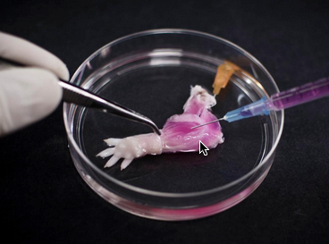
A suspension of muscle progenitor cells is injected into the cell-free matrix of a decellularized rat limb, which provides shape and structure onto which regenerated tissue can grow (credit: Bernhard Jank, MD, Ott Laboratory, Massachusetts General Hospital Center for Regenerative Medicine)

A team of Massachusetts General Hospital (MGH) investigators has made the first steps towards developing bioartificial replacement limbs suitable for transplantation.

In a Biomaterials journal report, the researchers describe using an experimental approach previously used to build bioartificial organs to engineer rat forelimbs with functioning vascular and muscle tissue. They also provided evidence that the same approach could be applied to the limbs of primates.

“The composite nature of our limbs makes building a functional biological replacement particularly challenging,” explains Harald Ott, MD, of the MGH Department of Surgery and the Center for Regenerative Medicine and assistant professor of Surgery at Harvard Medical School, senior author of the paper.

The progenitor cells needed to regenerate all of the tissues that make up a limb could be provided by the potential recipient. The problem is that limbs contain muscles, bone, cartilage, blood vessels, tendons, ligaments and nerves — each of which has to be rebuilt and requires a specific supporting structure (“matrix”), a step that has been a missing, he explained.

“We have shown that we can maintain the matrix of all of these tissues in their natural relationships to each other, that we can culture the entire construct over prolonged periods of time, and that we can repopulate the vascular system and musculature.”

Engineering a bioartificial limb

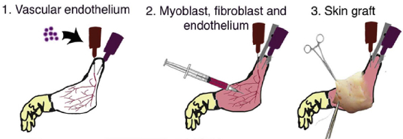
Procedure for composite tissue engineering. (1) Vascular endothelial cells are instilled into the vascular system of acellular composite tissue grafts. (2) Myoblasts, fibroblasts and endothelial cells are injected into the muscle compartment on day 2 of whole organ culture. (3) Full-thickness skin grafts are transplanted onto engineered constructs on day 10 of in vitro culture. (credit: B.J. Jank et al. / Biomaterials)

The current study uses technology Ott discovered as a research fellow at the University of Minnesota, in which living cells are stripped from a donor organ with a detergent solution and the remaining matrix is then repopulated with progenitor cells appropriate to the specific organ.

His team and others at MGH and elsewhere have used this decellularization technique to regenerate kidneyslivershearts, and lungs from animal models, but this is the first reported use to engineer the more complex tissues of a bioartificial limb.

The same decellularization process used in the whole-organ studies — perfusing a detergent solution through the vascular system — was used to strip all cellular materials from forelimbs removed from deceased rats in a way that preserved the primary vasculature and nerve matrix.

After thorough removal of cellular debris — a process that took a week — what remained was the cell-free matrix that provides structure to all of a limb’s composite tissues.  At the same time, populations of muscle and vascular cells were being grown in culture.

Bioreactor for growing a forelimb

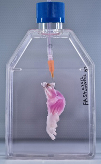
After vascular and muscle progenitors have been introduced into a decellularized rat limb, it is suspended in a bioreactor, which provides a nutrient solution and electrical stimulation to support and promote the growth of new tissues. (Bernhard Jank, MD, Ott Laboratory, Massachusetts General Hospital Center for Regenerative Medicine)

The research team then cultured the forelimb matrix in a bioreactor, within which vascular cells were injected into the limb’s main artery to regenerate veins and arteries.  Muscle progenitors were injected directly into the matrix sheaths that define the position of each muscle.

After five days in culture, electrical stimulation was applied to the potential limb graft to further promote muscle formation, and after two weeks, the grafts were removed from the bioreactor.

Analysis of the bioartificial limbs confirmed the presence of vascular cells along blood vessel walls and muscle cells aligned into appropriate fibers throughout the muscle matrix.

Functional testing of the isolated limbs showed that electrical stimulation of muscle fibers caused them to contract with a strength 80 percent of what would be seen in newborn animals.

The vascular systems of bioengineered forelimbs transplanted into recipient animals quickly filled with blood, which continued to circulate, and electrical stimulation of muscles within transplanted grafts flexed the wrists and digital joints of the animals’ paws.

The research team also successfully decellularized baboon forearms to confirm the feasibility of using this approach on the scale that would be required for human patients.

Replicating with human cells

Ott notes that, while regrowing nerves within a limb graft and reintegrating them into a recipient’s nervous system is one of the next challenges that needs to be faced, the experience of patients who have received hand transplants is promising.

“In clinical limb transplantation, nerves do grow back into the graft,  enabling both motion and sensation, and we have learned that this process is largely guided by the nerve matrix within the graft. We hope in future work to show that the same will apply to bioartificial grafts.

“Additional next steps will be replicating our success in muscle regeneration with human cells and expanding that to other tissue types, such as bone, cartilage and connective tissue.”

The study was supported by a New Innovator Award from the National Institutes of Health.

The authors note that more than 1.5 million individuals in the U.S. have lost a limb. Over the past two decades, a number of patients have received donor hand transplants, which also expose recipients to the risks of life-long immunosuppressive therapy.


Ott Laboratory | Rat Tissue Decellularization


Abstract of Engineered composite tissue as a bioartificial limb graft

The loss of an extremity is a disastrous injury with tremendous impact on a patient’s life. Current mechanical prostheses are technically highly sophisticated, but only partially replace physiologic function and aesthetic appearance. As a biologic alternative, approximately 70 patients have undergone allogeneic hand transplantation to date worldwide. While outcomes are favorable, risks and side effects of transplantation and long-term immunosuppression pose a significant ethical dilemma. An autologous, bio-artificial graft based on native extracellular matrix and patient derived cells could be produced on demand and would not require immunosuppression after transplantation. To create such a graft, we decellularized rat and primate forearms by detergent perfusion and yielded acellular scaffolds with preserved composite architecture. We then repopulated muscle and vasculature with cells of appropriate phenotypes, and matured the composite tissue in a perfusion bioreactor under electrical stimulation in vitro. After confirmation of composite tissue formation, we transplanted the resulting bio-composite grafts to confirm perfusion in vivo.

First multi-organ transplant that includes skull and scalp

James Boyson (credit: CNN)

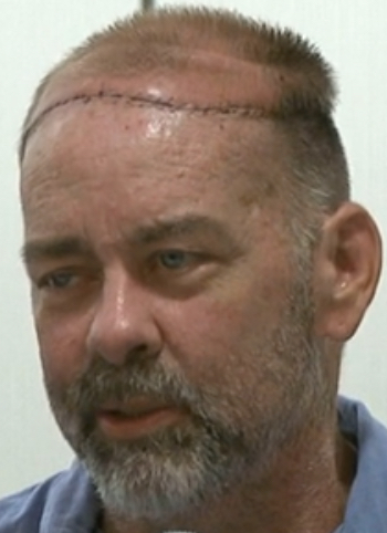
James Boysen, a 55-year-old software developer from Austin, Texas has become the first patient to receive a scalp and skull transplant while receiving kidney and pancreas transplants.

More than 50 health care professionals from Houston Methodist Hospital and The University of Texas MD Anderson Cancer Center assisted with or supported the double surgery over a period of more than 24 hours.

“This was a very complex surgery because we had to transplant the tissues utilizing microsurgery,” said Michael Klebuc, M.D., the surgeon who led the Houston Methodist Hospital Plastic Surgery Team.

“Imagine connecting blood vessels 1/16 of an inch under a microscope with tiny stitches about half the diameter of a human hair being done with tools that one would use to make a fine Swiss watch.”

In 2006, Boysen was diagnosed with leiomyosarcoma, a rare cancer of the smooth muscle, on his scalp. Successfully treated with chemotherapy and radiation, he was left with a large, deep wound on his head that included the scalp and the full thickness of his skull down to his brain.

In addition to the wound, which would require a major reconstructive undertaking, Boysen’s kidney and pancreas, which were first transplanted in 1992, were failing. Diagnosed with diabetes at age 5, Boysen’s declining condition over the years prompted the original double-organ transplant.

CNN video