Autistic brain is hyper-functional — needs predictable, paced environments, study finds

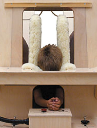
Part of the calming “Squeeze Machine” designed by Temple Grandin (credit: Therafin Corp.)

A new open-access study shows that social and sensory overstimulation drives autistic behaviors and supports the unconventional view that the autistic brain is actually hyper-functional. The research offers new hope, with therapeutic emphasis on paced and non-surprising environments tailored to the individual’s sensitivity.

For decades, autism has been viewed as a form of mental retardation, a brain disease that destroys children’s ability to learn, feel and empathize, thus leaving them disconnected from our complex and ever-changing social and sensory surroundings. From this perspective, the main kind of therapeutic intervention in autism to date aims at strongly engaging the child to revive brain functions believed dormant.

Predictability is key

Now researchers at the Swiss Federal Institute of Technology in Lausanne (EPFL) have completed a study that turns this traditional view of autism completely around. The study, conducted on rats exposed to a known risk factor in humans, demonstrates that unpredictable environmental stimulation drives autistic symptoms at least as much as an impoverished environment does.

It also shows that predictable stimulation can prevent these symptoms.

The study is also evidence for a drastic shift in the clinical approach to autism, away from the idea of a damaged brain that demands extensive stimulation. Instead, autistic brains may be hyper-functional and thus require enriched environments that are non-surprising, structured, safe, and tailored to a particular individual’s sensitivity.

“The valproate rat model used is highly relevant for understanding autism, because children exposed to valproate in the womb have an increased chance of presenting autism after birth,” says Prof. Henry Markram, co-author of the study and father of a child with autism. He notes that the rats exposed to valproate in early embryonic development demonstrate behavioral, anatomical and neurochemical abnormalities that are comparable to characteristics of human autism.

The scientists here show that if these rats are reared in a home environment that is calm, safe, and highly predictable with little surprise — while still rich in sensory and social engagement — they do not develop symptoms of emotional over-reactivity such as fear and anxiety, nor social withdrawal or sensory abnormalities.

“We were amazed to see that environments lacking predictability, even if enriched, favored the development of hyper-emotionality in rats exposed to the prenatal autism risk factor,” says Markram.

The study critically shows that in certain individuals, non-predictable environments lead to the development of a wider range of negative symptoms, including social withdrawal and sensory abnormalities. Such symptoms normally prevent individuals from fully benefiting from and contributing to their surroundings, and are thus the targets of therapeutic success.

The study identifies drastically opposite behavioral outcomes depending on levels of predictability in the enriched environment, and suggests that the autistic brain is unusually sensitive to predictability in rearing environment, but to different extent in different individuals.

Hyper-functional brain microcircuits

The study is strong evidence for the Intense World Theory of Autism, proposed in 2007 by neuroscientists Kamila Markram and Henry Markram, both co-authors on the present study. This theory is based on recent research suggesting that the autistic brain, in both humans and animal models, reacts differently to stimuli.

It proposes that an interaction — between an individual’s genetic background with biologically toxic events early in embryonic development — triggers a cascade of abnormalities that create hyper-functional brain microcircuits, the functional units of the brain.

Once activated, these hyper-functional circuits could become autonomous and affect further brain functional connectivity and development. These would lead to an experience of the world as intense, fragmented, and overwhelming; while differences in severity between persons with autism would stem from the system affected and the timing of the effect.

Stable, structured environment

Instead, a stable, structured environment rich in stimuli could help children with autism, by providing a safe haven from an overload of sensory and emotional stimuli, the authors suggest.

This study has immediate implications for clinical and research settings. It suggests that if brain hyper-function can be diagnosed soon after birth, at least some of the debilitating effects of a supercharged brain can be prevented by highly specialized environmental stimulation that is safe, consistent, controlled, announced and only changed very gradually at the pace determined by each child.

The research supports the work of Temple Grandin, PhD, an author and professor of animal science at Colorado State University. One of the therapeutic methods she developed (and used herself) was the “hug machine” (AKA “squeeze machine”), a deep-pressure device designed to calm hypersensitive persons. The device is featured in an award-winning biographical film, Temple Grandin.


Abstract of Predictable enriched environment prevents development of hyper-emotionality in the VPA rat model of autism

Understanding the effects of environmental stimulation in autism can improve therapeutic interventions against debilitating sensory overload, social withdrawal, fear and anxiety. Here, we evaluate the role of environmental predictability on behavior and protein expression, and inter-individual differences, in the valproic acid (VPA) model of autism. Male rats embryonically exposed (E11.5) either to VPA, a known autism risk factor in humans, or to saline, were housed from weaning into adulthood in a standard laboratory environment, an unpredictably enriched environment, or a predictably enriched environment. Animals were tested for sociability, nociception, stereotypy, fear conditioning and anxiety, and for tissue content of glutamate signaling proteins in the primary somatosensory cortex, hippocampus and amygdala, and of corticosterone in plasma, amygdala and hippocampus. Standard group analyses on separate measures were complemented with a composite emotionality score, using Cronbach’s Alpha analysis, and with multivariate profiling of individual animals, using Hierarchical Cluster Analysis. We found that predictable environmental enrichment prevented the development of hyper-emotionality in the VPA-exposed group, while unpredictable enrichment did not. Individual variation in the severity of the autistic-like symptoms (fear, anxiety, social withdrawal and sensory abnormalities) correlated with neurochemical profiles, and predicted their responsiveness to predictability in the environment. In controls, the association between socio-affective behaviors, neurochemical profiles and environmental predictability was negligible. This study suggests that rearing in a predictable environment prevents the development of hyper-emotional features in animals exposed to an autism risk factor, and demonstrates that unpredictable environments can lead to negative outcomes, even in the presence of environmental enrichment.

Improving the experience of the audience with digital instruments

Virtual content being displayed on stage and overlapping the instruments and the performers (credit: Florent Berthaut)

University of Bristol researchers have developed a new augmented-reality display that allows audiences to better appreciate digital musical performances

The research team from the University’s Bristol Interaction and Graphics (BIG) has been investigating how to improve the audiences experience during performances with digital musical instruments, which are played by manipulating buttons, mich, and various other controls.

Funded by a Marie Curie grant, the IXMI project, led by Florent Berthaut, aims to show the mechanisms of digital instruments, using 3D virtual content and mixed-reality displays.

Their first creation Reflets is a mixed-reality environment that allows for displaying virtual content anywhere on stage, even overlapping the instruments or the performers. It does not require the audience to wear glasses or to use their smartphones to see the augmentations, which remain consistent at all positions in the audience.

Reflets relies on combining the audience and stage spaces using reflective transparent surfaces and having the audience and performers reveal the virtual content by intersecting it with their bodies or physical props.

The research is being presented at the 15th International Conference on New Interfaces for Musical Expression (NIME) in the U.S. [May 31 -- June 3].


BristolIG | Ixmi: Improving the experience of the audience with digital instruments

Missing link found between brain, immune system

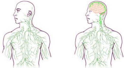
Maps of the lymphatic system: old (left) and updated (right) (credit: University of Virginia Health System)

Overrturning decades of textbook teaching, researchers at the University of Virginia School of Medicine have discovered that the brain is directly connected to the immune system by vessels previously thought not to exist.

The finding could have significant implications for the study and treatment of neurological diseases ranging from autism to Alzheimer’s disease to multiple sclerosis.

“It changes entirely the way we perceive the neuro-immune interaction. We always perceived it before as something esoteric that can’t be studied. But now we can ask mechanistic questions.” said Jonathan Kipnis, PhD, professor in the UVA Department of Neuroscience and director of UVA’s Center for Brain Immunology and Glia (BIG).

“We believe that for every neurological disease that has an immune component to it, these vessels may play a major role,” Kipnis said. “Hard to imagine that these vessels would not be involved in a [neurological] disease with an immune component.”

“Very Well Hidden”

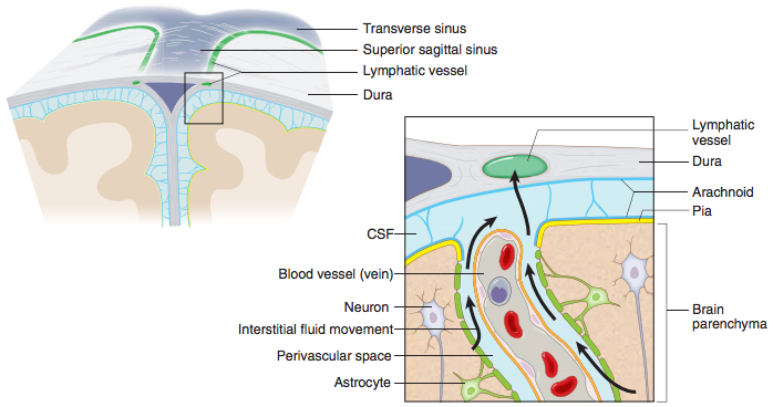
A schematic representation of a connection between the glymphatic system, responsible for collecting of the interstitial fluids from within the central nervous system parenchyma to cerebrospinal fluid, and the newly identified meningeal lymphatic vessels (credit: Antoine Louveau et al./Nature)

The discovery was made possible by the work of Antoine Louveau, PhD, a postdoctoral fellow in Kipnis’ lab, who noticed vessel-like patterns in the distribution of immune cells on his slides of a mouse’s meninges — the membranes covering the brain.

So how did the brain’s lymphatic vessels manage to escape notice all this time? Kipnis described them as “very well hidden” — they follow a major blood vessel down into the sinuses, an area difficult to image. “It’s so close to the blood vessel, you just miss it… if you don’t know what you’re after.”

Alzheimer’s, Autism, MS and Beyond

The unexpected presence of the lymphatic vessels raises a tremendous number of questions that now need answers, both about the workings of the brain and the diseases that plague it.

For example: “In Alzheimer’s, there are accumulations of big protein chunks in the brain,” Kipnis said. “We think they may be accumulating in the brain because they’re not being efficiently removed by these vessels.”

He noted that the vessels look different with age, so the role they play in aging is another avenue to explore. And there’s an enormous array of other neurological diseases, from autism to multiple sclerosis, that must be reconsidered in light of the presence of something science insisted did not exist.

The findings have been published online by the journal Nature and will appear in a forthcoming print edition.


Abstract of Structural and functional features of central nervous system lymphatic vessels

One of the characteristics of the central nervous system is the lack of a classical lymphatic drainage system. Although it is now accepted that the central nervous system undergoes constant immune surveillance that takes place within the meningeal compartment123, the mechanisms governing the entrance and exit of immune cells from the central nervous system remain poorly understood456. In searching for T-cell gateways into and out of the meninges, we discovered functional lymphatic vessels lining the dural sinuses. These structures express all of the molecular hallmarks of lymphatic endothelial cells, are able to carry both fluid and immune cells from the cerebrospinal fluid, and are connected to the deep cervical lymph nodes. The unique location of these vessels may have impeded their discovery to date, thereby contributing to the long-held concept of the absence of lymphatic vasculature in the central nervous system. The discovery of the central nervous system lymphatic system may call for a reassessment of basic assumptions in neuroimmunology and sheds new light on the aetiology of neuroinflammatory and neurodegenerative diseases associated with immune system dysfunction.

Robot servants push the boundaries in HUMANS

(credit: AMC)

AMC announced today HUMANS, an eight-part TV science-fiction thriller that takes place in a parallel present featuring sophisticated, life-like robot servants and caregivers called Synths (personal synthetics).

The show explores conflicts as the lines between humans and machines become increasingly blurred.

The upgrade you’ve been waiting for is here (credit: AMC)

The series is set to premiere on AMC June 28 with HUMANS 101: The Hawkins family buys a Synth, Anita. But are they in danger from this machine and the young man Leo who seems desperate to find her?

The show features Oscar-winning actor William Hurt, Katherine Parkinson (The IT Crowd), Colin Morgan (Merlin), and Gemma Chan (Secret Diary of a Call Girl).


AMC

More HUMANS trailers

Emulating animals, these robots can recover from damage in two minutes

Researchers in France and the U.S. have developed a new technology that enables robots to quickly recover from an injury in less than two minutes, similar to how injured animals adapt. Such autonomous mobile robots would be useful in remote or hostile environments such as disaster areas, space, and deep oceans.

The video above shows a six-legged robot that adapts to keep walking even if two of its legs are broken. It also shows a robotic arm that learned how to correctly place an object even with several broken motors.

“When injured, animals do not start learning from scratch,” says Jean-Baptiste Mouret from Pierre and Marie Curie University. “Instead, they have intuitions about different ways to behave. These intuitions allow them to intelligently select a few, different behaviors to try out and, after these tests, they choose one that works in spite of the injury. We made robots that can do the same.”

The researchers developed an “Intelligent Trial and Error” algorithm that allows robots to emulate animals: the robots conduct experiments to rapidly discover a compensatory behavior that works despite the damage.

“For example, if walking, mostly on its hind legs, does not work well, it will next try walking mostly on its front legs,”  explains Antoine Cully, lead author of a May 28 cover article on this research in the journal Nature. “What’s surprising is how quickly it can learn a new way to walk. It’s amazing to watch a robot go from crippled and flailing around to efficiently limping away in about two minutes.”


Abstract of Robots that can adapt like animals.

Robots have transformed many industries, most notably manufacturing1, and have the power to deliver tremendous benefits to society, such as in search and rescue2, disaster response3, health care4 and transportation5. They are also invaluable tools for scientific exploration in environments inaccessible to humans, from distant planets6 to deep oceans7. A major obstacle to their widespread adoption in more complex environments outside factories is their fragility68. Whereas animals can quickly adapt to injuries, current robots cannot ‘think outside the box’ to find a compensatory behaviour when they are damaged: they are limited to their pre-specified self-sensing abilities, can diagnose only anticipated failure modes9, and require a pre-programmed contingency plan for every type of potential damage, an impracticality for complex robots68. A promising approach to reducing robot fragility involves having robots learn appropriate behaviours in response to damage1011, but current techniques are slow even with small, constrained search spaces12. Here we introduce an intelligent trial-and-error algorithm that allows robots to adapt to damage in less than two minutes in large search spaces without requiring self-diagnosis or pre-specified contingency plans. Before the robot is deployed, it uses a novel technique to create a detailed map of the space of high-performing behaviours. This map represents the robot’s prior knowledge about what behaviours it can perform and their value. When the robot is damaged, it uses this prior knowledge to guide a trial-and-error learning algorithm that conducts intelligent experiments to rapidly discover a behaviour that compensates for the damage. Experiments reveal successful adaptations for a legged robot injured in five different ways, including damaged, broken, and missing legs, and for a robotic arm with joints broken in 14 different ways. This new algorithm will enable more robust, effective, autonomous robots, and may shed light on the principles that animals use to adapt to injury.

MIT cheetah robot now jumps over obstacles autonomously


Massachusetts Institute of Technology| MIT cheetah robot lands the running jump

The MIT researchers who built a robotic cheetah have now trained it to see and jump over hurdles as it runs — making it the first four-legged robot to run and jump over obstacles autonomously.

The robot estimates an obstacle’s height and distance, gauges the best distance from which to jump, and adjusts its stride to land just short of the obstacle, before exerting enough force to push up and over. Based on the obstacle’s height, the robot then applies a certain amount of force to land safely, before resuming its initial pace.

In experiments on a treadmill and an indoor track, the cheetah robot successfully cleared obstacles up to 18 inches tall — more than half of the robot’s own height — while maintaining an average running speed of 5 miles per hour.

“A running jump is a truly dynamic behavior,” says Sangbae Kim, an assistant professor of mechanical engineering at MIT. “You have to manage balance and energy, and be able to handle impact after landing. Our robot is specifically designed for those highly dynamic behaviors.”

Onboard LIDAR + path-planing algorithm –> autonomous control

As KurzweilAI reported last September, the engineers previously demonstrated that the robotic cheetah was able to run untethered— performed “blind,” without the use of cameras or other vision systems.

Now, the robot can “see,” with the use of onboard LIDAR — a visual system that uses reflections from a laser to map terrain (also used in autonomous vehicles). The team developed a three-part algorithm to plan out the robot’s path, based on LIDAR data. Both the vision and path-planning system are onboard the robot, giving it complete autonomous control.

The team tested the cheetah’s jumping ability first on a treadmill, then on a track. On the treadmill, the robot ran tethered in place, as researchers placed obstacles of varying heights on the belt. After multiple runs, the robot successfully cleared about 70 percent of the hurdles.

In comparison, tests on an indoor track proved much easier, as the robot had more space and time in which to see, approach, and clear obstacles. In these runs, the robot successfully cleared about 90 percent of obstacles.

The team is now working on getting the MIT cheetah to jump over hurdles while running on softer terrain, like a grassy field.

This research was funded in part by the Defense Advanced Research Projects Agency .

Scientists recover ‘lost’ memories using brain stimulation by blue light

(credit: Christine Daniloff/MIT)

MIT researchers have found they were able to reactivate memories in mice that could not otherwise be retrieved, using optogenetics — in which proteins are added to neurons to allow them to be activated with light.

The breakthrough finding, in a paper published Thursday (May 28) in the journal Science, appears to answer a longstanding question in neuroscience regarding amnesia.

Damaged or blocked memory?

Neuroscience researchers have for many years debated whether retrograde amnesia — which follows traumatic injury, stress, or diseases such as Alzheimer’s — is caused by damage to specific brain cells, meaning a memory cannot be stored, or if access to that memory is somehow blocked, preventing its recall.

The answer, according to Susumu Tonegawa, the Picower Professor in MIT’s Department of Biology and director of the RIKEN-MIT Center at the Picower Institute for Learning and Memory: “Amnesia is a problem of retrieval impairment.”

Memory researchers have previously speculated that somewhere in the brain network is a population of neurons that are activated during the process of acquiring a memory, causing enduring physical or chemical changes.

If these groups of neurons are subsequently reactivated by a trigger such as a particular sight or smell, for example, the entire memory is recalled. These neurons are known as “memory engram cells.”

Blocking, then activating memories with light

Until now, no one has been able to show that these groups of neurons undergo enduring chemical changes, in a process known as memory consolidation. One such change, known as “long-term potentiation” (LTP), involves the strengthening of synapses, the structures that allow groups of neurons to send signals to each other, as a result of learning and experience.

To find out if these chemical changes do indeed take place, the researchers first identified a group of engram cells in the hippocampus that, when activated using optogenetic tools, were able to express a memory.

When they then recorded the activity of this particular group of cells, they found that the synapses connecting them had been strengthened. “We were able to demonstrate for the first time that these specific cells — a small group of cells in the hippocampus — had undergone this augmentation of synaptic strength,” Tonegawa says.

The researchers then attempted to discover what happens to memories without this consolidation process. By administering a compound called anisomycin, which blocks protein synthesis within neurons, immediately after mice had formed a new memory, the researchers were able to prevent the synapses from strengthening.

When they returned one day later and attempted to reactivate the memory using an emotional trigger, they could find no trace of it. “So even though the engram cells are there, without protein synthesis those cell synapses are not strengthened, and the memory is lost,” Tonegawa says.

But startlingly, when the researchers then reactivated the protein synthesis-blocked engram cells using optogenetic tools, they found that the mice exhibited all the signs of recalling the memory in full.

“If you test memory recall with natural recall triggers in an anisomycin-treated animal, it will be amnesiac, you cannot induce memory recall,” Tonegawa says. “But if you go directly to the putative engram-bearing cells and activate them with light, you can restore the memory, despite the fact that there has been no LTP.”

Memories are stored in a circuit of groups of cells in multiple brain areas, not synapses

Further studies carried out by Tonegawa’s group demonstrated that memories are stored not in synapses strengthened by protein synthesis in individual engram cells, but in a circuit, or “pathway” of multiple groups of engram cells and the connections between them.

“We are proposing a new concept, in which there is an engram cell ensemble pathway, or circuit, for each memory,” he says. “This circuit encompasses multiple brain areas and the engram cell ensembles in these areas are connected specifically for a particular memory.”

The research dissociates the mechanisms used in memory storage from those of memory retrieval, according to Ryan. “The strengthening of engram synapses is crucial for the brain’s ability to access or retrieve those specific memories, while the connectivity pathways between engram cells allows the encoding and storage of the memory information itself,” he says.

Changes in synaptic strength and in spine properties have long been associated with learning and memory, according to Alcino Silva, director of the Integrative Center for Learning and Memory at the University of California at Los Angeles.

“This groundbreaking paper suggests that these changes may not be as critical for memory as once thought, since under certain conditions, it seems to be possible to disrupt these changes and still preserve memory,” he says. “Instead, it appears that these changes may be needed for memory retrieval, a mysterious process that has so far evaded neuroscientists.”


Abstract of Engram cells retain memory under retrograde amnesia

Memory consolidation is the process by which a newly formed and unstable memory transforms into a stable long-term memory. It is unknown whether the process of memory consolidation occurs exclusively through the stabilization of memory engrams. By using learning-dependent cell labeling, we identified an increase of synaptic strength and dendritic spine density specifically in consolidated memory engram cells. Although these properties are lacking in engram cells under protein synthesis inhibitor–induced amnesia, direct optogenetic activation of these cells results in memory retrieval, and this correlates with retained engram cell–specific connectivity. We propose that a specific pattern of connectivity of engram cells may be crucial for memory information storage and that strengthened synapses in these cells critically contribute to the memory retrieval process.

Light electric stimulation of the brain may improve memory for people with schizophrenia

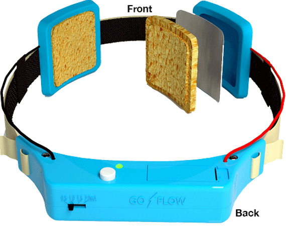
Transcranial direct-current stimulation device (credit: GoFlow)

Lightly stimulating the brain with transcranial direct current stimulation (tDCS) may improve short-term memory in people with schizophrenia, according to a new study by researchers at the Johns Hopkins University School of Medicine.

The tDCS procedure involves placing sponge-covered electrodes on the head and passing a weak electrical current between them.

David Schretlen, Ph.D., a professor of psychiatry and behavioral sciences at the Johns Hopkins University School of Medicine, reasoned that this type of brain stimulation might ease some of the cognitive difficulties that afflict people with schizophrenia.

A test based on prefrontal cortex stimulation

To test that possibility, Schretlen and Johns Hopkins colleagues targeted a brain region called the left dorsolateral prefrontal cortex, which plays an important role in short-term or working memory and is abnormal in people with schizophrenia, according to Schretlen.

Schretlen recruited 11 participants: five adults with confirmed schizophrenia and six of their close relatives (parents, siblings, and children of people with schizophrenia show some of the same abnormalities to a lesser degree, says Schretlen).

Each participant received two 30-minute treatments — one using a negative electrical charge, which the researchers thought might prove beneficial — and the other using a positive charge as a control. During and after each treatment, participants completed a battery of cognitive tests.

Thinking improvements

There were two notable results:

  • On tests of verbal and visual working memory, participants performed significantly better after receiving a negative charge, and the effects were “surprisingly strong,” says Schretlen.
  • Participants did better at the challenging task of switching between naming categories of items in a supermarket after a negatively charged treatment. The stimulation “was associated with better performance on working memory and subtle changes in word retrieval,” Schretlen says. People with schizophrenia often struggle to find the right words, he says. Because the prefrontal cortex contains a brain region responsible for word retrieval, Schretlen reasoned that transcranial direct current stimulation might help.

Schretlen is now studying transcranial direct current stimulation in a larger sample of patients using repeated sessions of stimulation, which he hopes will induce lasting benefits.

“Cognitive impairment is as ubiquitous as hallucinations in schizophrenia, yet medications only treat the hallucinations,” Schretlen says. “So even with medication, affected individuals often remain very disabled.” His hope is that transcranial direct current stimulation could give people with schizophrenia a shot at leading a more normal life.

Other findings

A related study last year showed that tDCS improved correction of mistakes. But another recent study found that after a repeated IQ test (which is normally expected to show improvements), IQ scores of people who underwent tDCS brain stimulation improved markedly less than did the IQ scores of people in the placebo group.

The tDCS procedure is also being studied by other researchers as a treatment for depression and Alzheimer’s-related memory loss, and to enhance recovery following strokes.

The research is described in a paper published online in Clinical Schizophrenia and Related Psychoses. The study was funded by the Therapeutic Cognitive Neuroscience Professorship; the Therapeutic Cognitive Neuroscience Fund; the Benjamin and Adith Miller Family Endowment on Aging, Alzheimer’s and Autism; and the National Institute on Child and Human Development.


Abstract of Can Transcranial Direct Current Stimulation Improve Cognitive Functioning in Adults with Schizophrenia?

Cognitive impairment is nearly ubiquitous in schizophrenia. First-degree relatives of persons with schizophrenia often show similar but milder deficits. Current methods for the treatment of schizophrenia are often ineffective in cognitive remediation. Since transcranial direct current stimulation (tDCS) can enhance cognitive functioning in healthy adults, it might provide a viable option to enhance cognition in schizophrenia. We sought to explore whether tDCS can be tolerated by persons with schizophrenia and potentially improve their cognitive functioning. We examined the effects of anodal versus cathodal tDCS on working memory and other cognitive tasks in five outpatients with schizophrenia and six first-degree relatives of persons with schizophrenia. Each participant completed tasks thought to be mediated by the prefrontal cortex during two 30-minute sessions of tDCS to the left and right dorsolateral prefrontal cortex (DLPFC). Anodal stimulation over the left DLPFC improved performance relative to cathodal stimulation on measures of working memory and aspects of verbal fluency relevant to word retrieval. The patient group showed differential changes in novel design production without alteration of overall productivity, suggesting that tDCS might be capable of altering selfmonitoring and executive control. All participants tolerated tDCS well. None withdrew from the study or experienced any adverse reaction. We conclude that adults with schizophrenia can tolerate tDCS while engaging in cognitive tasks and that tDCS can alter their performance.

A 99% biodegradable computer chip

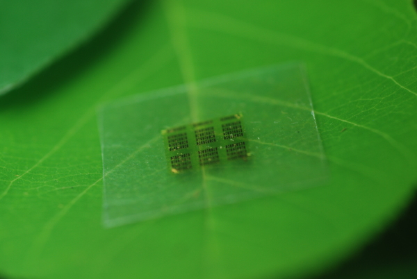
A cellulose nanofibril (CNF) computer chip shown on a leaf (credit: Yei Hwan Jung, Wisconsin Nano Engineering Device Laboratory)

University of Wisconsin-Madison and U.S. Department of Agriculture Forest Products Laboratory (FPL) researchers have jointly developed a wood chip in an effort to alleviate the environmental burden* of electronic devices.

Well, actually, a wood-substrate-based semiconductor chip. They replaced the silicon substrate portion in a conventional chip with environment-friendly cellulose nanofibril (CNF). CNF is a flexible, biodegradable material made from wood, as the researchers note in an open-access paper published May 26 in the journal Nature Communications.

“[More than 99%] of the material in a chip is support,” said Zhiyong Cai, project leader of an engineering composite science research group at FPL. With the new substrate, the chips are “so safe you can put them in the forest and fungus will degrade it. They become as safe as fertilizer.”

The new material is especially important for microwave chips (such as those used in mobile phones) made with gallium arsenide, which is especially difficult to fabricate on foreign substrates. That’s because of the small feature sizes and high temperature processes required for high performance.

Cai’s group addressed two key barriers to using wood-derived materials in an electronics setting: surface roughness and thermal expansion. “You don’t want it to expand or shrink too much. Wood is a natural hydroscopic [water-absorbing] material and could attract moisture from the air and expand,” Cai says. “With an epoxy coating on the surface of the CNF, we solved both [problems].”

* In 2007, it was estimated that over 426,000 cell phones (most of them were still functional) and 112,000 computers were discarded every day in the US, totalling 3.2 million tons of electronic waste generated per year, the researcher note in the paper.


Abstract of High-performance green flexible electronics based on biodegradable cellulose nanofibril paper

Today’s consumer electronics, such as cell phones, tablets and other portable electronic devices, are typically made of non-renewable, non-biodegradable, and sometimes potentially toxic (for example, gallium arsenide) materials. These consumer electronics are frequently upgraded or discarded, leading to serious environmental contamination. Thus, electronic systems consisting of renewable and biodegradable materials and minimal amount of potentially toxic materials are desirable. Here we report high-performance flexible microwave and digital electronics that consume the smallest amount of potentially toxic materials on biobased, biodegradable and flexible cellulose nanofibril papers. Furthermore, we demonstrate gallium arsenide microwave devices, the consumer wireless workhorse, in a transferrable thin-film form. Successful fabrication of key electrical components on the flexible cellulose nanofibril paper with comparable performance to their rigid counterparts and clear demonstration of fungal biodegradation of the cellulose-nanofibril-based electronics suggest that it is feasible to fabricate high-performance flexible electronics using ecofriendly materials.

Psychedelic drugs should be legally reclassified, says psychiatrist

The Persistence of Memory (credit: Salvador Dali)

Psychedelic drugs such as LSD are much less harmful than claimed and should be legally reclassified to allow further research on their medical use, says James Rucker, a psychiatrist and honorary lecturer at the Institute of Psychiatry, Psychology and Neuroscience, King’s College London.

These substances “were extensively used and researched in clinical psychiatry” before their prohibition in 1967 and many trials of these drugs in the 1950s and 1960s suggested “beneficial change in many psychiatric disorders.”

Nonetheless, in the UK, psychedelic drugs were legally classified as schedule 1 class A drugs; that is, as having “no accepted medical use and the greatest potential for harm, despite the research evidence to the contrary,” he writes.

Clinical efficacy shown in anxiety, obsessive compulsive disorder, addiction, and headaches

Rucker makes these points:

  • Psychedelics remain more legally restricted than heroin and cocaine. “But no evidence indicates that psychedelic drugs are habit forming; little evidence indicates that they are harmful in controlled settings; and much historical evidence shows that they could have use in common psychiatric disorders.” In fact, recent studies indicate that psychedelics have “clinical efficacy in anxiety associated with advanced cancer, obsessive compulsive disorder, tobacco and alcohol addiction, and cluster headaches,” he writes.
  • At present, larger clinical studies on psychedelics are made “almost impossible by the practical, financial and bureaucratic obstacles” imposed by their schedule 1 classification. Currently, only one manufacturer in the world produces psilocybin for trial purposes, he says, at a “prohibitive” cost of £100,000 for 1 g (50 doses).
  • In the UK, to hold a schedule 1 drug, institutions require a license, which costs about £5,000, he adds. Only four hospitals currently hold such licenses, which come with regular police or home office inspections and onerous rules on storage and transport, so “clinical research using psychedelics costs 5–10 times that of research into less restricted (but more harmful) drugs such as heroin.” As a result, “almost all grant funders are uncomfortable funding research into psychedelics,” while prohibition as a condition of UN membership is “arguably causing more harm than it prevents.”
  • Psychedelics are neither harmful nor addictive compared with other controlled substances. He calls on the UK Advisory Council on the Misuse of Drugs and the 2016 UN General Assembly Special Session on Drugs, “to recommend that psychedelics be reclassified as schedule 2 compounds to enable a comprehensive, evidence based assessment of their therapeutic potential.”


The Beatles — A Day in the Life