Researchers can sequence DNA from airplane toilets to trace resistance genes and potential pathogens.
The post Airplane Poop Could Help Track Global Disease Outbreaks appeared first on WIRED.

Science and reality

Researchers can sequence DNA from airplane toilets to trace resistance genes and potential pathogens.
The post Airplane Poop Could Help Track Global Disease Outbreaks appeared first on WIRED.

The best data medical researchers have on radiation risk comes from long-term studies of the survivors of Hiroshima and Nagasaki. But they might not be enough.
The post How Hiroshima Survivors Are Leaving A Legacy For Science appeared first on WIRED.

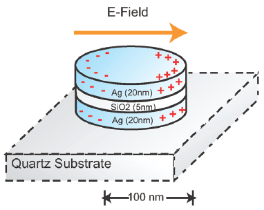
A nanoplasmonic resonator (NPR) consists of a thin silicon dioxide layer sandwiched between metallic nanodisks. NPRs can enhance surface-enhanced Raman spectroscopic (SERS) signals by a factor of 60 billion to detect target molecules with high sensitivity. (credit: Cheng Sun et al./ ACS Nano)
Imagine being able to test your food in your kitchen to quickly determine if it carried any deadly microbes. Technology now being commercialized by Optokey may soon make that possible.
Optokey, a startup based in Hayward, California, has developed a miniaturized sensor using surface-enhanced Raman spectroscopy (SERS) that can quickly and accurately detect or diagnose substances at a molecular level. The technology is based on research conducted at Lawrence Berkeley National Laboratory (Berkeley Lab) and published in 2010.
Molecular fingerprinting
“Our system can do chemistry, biology, biochemistry, molecular biology, clinical diagnosis, and chemical analysis,” said Optokey president and co-founder Fanqing Frank Chen, a scientist at Berkeley Lab who was co-author of an ACS Nano paper on the research. The system can be implemented “very cheaply, without much human intervention,” he said.
SERS is a highly sensitive analytical tool used for “molecular fingerprinting,” but the results have not easily reproducible. Chen and colleagues developed a solution to this problem using what they called “nanoplasmonic resonators,” which measures the interaction of photons with an activated surface using nanostructures to do chemical and biological sensing. The method produces measurements much more reliably.
“At Optokey we’re able to mass produce this nanoplasmonic resonator on a wafer scale,” Chen said. “We took something from the R&D realm and turned it into something industrial-strength.”
The miniaturized sensors use a microfluidic control system for “lab on a chip” automated liquid sampling. “We’re leveraging knowledge acquired from high-tech semiconductor manufacturing methods to get the cost, the volume, and the accuracy in the chip,” said VP of Manufacturing Robert Chebi, a veteran of the microelectronic industry who previously worked at Lam Research and Applied Materials. “We’re also leveraging all the knowledge in lasers and optics for this specific Raman-based method.”
A biochemical nose
Chebi calls Optokey’s product a “biochemical nose,” or an advanced nanophotonic automated system, with sensitivity to the level of a single molecule, far superior to sensors on the market today, he claims. “Today’s detection and diagnosis methods are far from perfect … Also, our system can provide information in minutes, or even on a continuous basis, versus other methods where it could take hours or even days, if samples have to be sent to another lab.”
The potential applications include food safety, environmental monitoring (of both liquids and gases), medical diagnosis, and chemical analysis. Optokey’s customers include a major European company interested in food safety, a Chinese petrochemical company interested in detecting impurities in its products, and a German company interested in point-of-care diagnosis.
“The product we’re envisioning is something that is compact and automated but also connected, and it can go into schools, restaurants, factories, hospitals, ambulances, airports, and even battlefields,” Chen said. Next, they plan to introduce it in the smart home, where a nanophotonic sensor could be built to scan for pollutants not just in food but also in air and water.
Key discovery: nanoplasmonic resonators
Ultimately, Chen and his Berkeley Lab group developed about 20 patents involving hybrid bionanomaterials. The key discovery that led to the formation of Optokey was the development of nanoplasmonic resonators to dramatically improve the signal and reliability of Raman spectroscopy. The method was initially used in the research lab to quickly and accurately detect a biomarker for prostate cancer, which has a high rate of false positives using conventional diagnostic tools.
“There was 10 years of research that went into this, funded by NIH, DARPA, the federal government, private foundations,” said Chen. “Berkeley Lab has a really good culture of multidisciplinary research, excellent engineering, and very strong basic science. Plus it has strong support for startups.”
Abstract of Time-Resolved Single-Step Protease Activity Quantification Using Nanoplasmonic Resonator Sensors
Protease activity measurement has broad application in drug screening, diagnosis and disease staging, and molecular profiling. However, conventional immunopeptidemetric assays (IMPA) exhibit low fluorescence signal-to-noise ratios, preventing reliable measurements at lower concentrations in the clinically important picomolar to nanomolar range. Here, we demonstrated a highly sensitive measurement of protease activity using a nanoplasmonic resonator (NPR). NPRs enhance Raman signals by 6.1 × 10¹⁰ times in a highly reproducible manner, enabling fast detection of proteolytically active prostate-specific antigen (paPSA) activities in real-time, at a sensitivity level of 6 pM (0.2 ng/mL) with a dynamic range of 3 orders of magnitude. Experiments on extracellular fluid (ECF) from the paPSA-positive cells demonstrate specific detection in a complex biofluid background. This method offers a fast, sensitive, accurate, and one-step approach to detect the proteases’ activities in very small sample volumes.
Eating a group of specific foods — known as the MIND diet — may slow cognitive decline among aging adults, even when the person is not at risk of developing Alzheimer’s disease, according to researchers at Rush University Medical Center.
This finding supplements a previous study by the research team, reported by KurzweiliAI in March, that found that the MIND diet may reduce a person’s risk in developing Alzheimer’s disease.
The researchers’ new study shows that older adults who followed the MIND diet more rigorously showed an equivalent of being 7.5 years younger cognitively than those who followed the diet least. Results of the study were recently published online in the journal Alzheimer’s & Dementia: The Journal of the Alzheimer’s Association.
So what is the MIND diet?
The MIND diet, which is short for “Mediterranean-DASH Diet Intervention for Neurodegenerative Delay,” was developed by Martha Clare Morris, ScD, a nutritional epidemiologist, and her colleagues. As the name suggests, the MIND diet is a hybrid of the Mediterranean and DASH (Dietary Approaches to Stop Hypertension) diets. Both have been found to reduce the risk of cardiovascular conditions, like hypertension, heart attack and stroke.
“Everyone experiences decline with aging; and Alzheimer’s disease is now the sixth leading cause of death in the U.S., which accounts for 60 to 80 percent of dementia cases. Therefore, prevention of cognitive decline, the defining feature of dementia, is now more important than ever,” Morris says. “Delaying dementia’s onset by just five years can reduce the cost and prevalence by nearly half.”
The MIND diet has 15 dietary components, including 10 “brain-healthy food groups” and five “unhealthy groups” to avoid — red meat, butter and stick margarine, cheese, pastries and sweets, and fried or fast food.
To adhere to and benefit from the MIND diet, a person would need to eat at least three servings of whole grains, a green leafy vegetable and one other vegetable every day — along with a glass of wine (or red-grape juice) — snack most days on nuts, have beans every other day or so, eat poultry and berries at least twice a week and fish at least once a week.
In addition, the study found that to have a real shot at avoiding the devastating effects of cognitive decline, he or she must limit intake of the designated unhealthy foods, especially butter (less than 1 tablespoon a day), sweets and pastries, whole fat cheese, and fried or fast food (less than a serving a week for any of the three).
Berries are the only fruit specifically to be included in the MIND diet. “Blueberries are one of the more potent foods in terms of protecting the brain,” Morris says, and strawberries also have performed well in past studies of the effect of food on cognitive function.
The National Institute of Aging-funded study evaluated cognitive change over a period of 4.7 years among 960 older adults who were free of dementia on enrollment. Averaging 81.4 years in age, the study participants also were part of the Rush Memory and Aging Project, a study of residents of more than 40 retirement communities and senior public housing units in the Chicago area.
During the course of the study, they received annual, standardized testing for cognitive ability in five areas — episodic memory, working memory, semantic memory, visuospatial ability and perceptual speed. The study group also completed annual food frequency questionnaires, allowing the researchers to compare participants’ reported adherence to the MIND diet with changes in their cognitive abilities as measured by the tests.
Abstract of MIND diet slows cognitive decline with aging
Background: The Mediterranean and dash diets have been shown to slow cognitive decline; however, neither diet is specific to the nutrition literature on dementia prevention.
Methods: We devised the Mediterranean-Dietary Approach to Systolic Hypertension (DASH) diet intervention for neurodegenerative delay (MIND) diet score that specifically captures dietary components shown to be neuroprotective and related it to change in cognition over an average 4.7 years among 960 participants of the Memory and Aging Project.
Results: In adjusted mixed models, the MIND score was positively associated with slower decline in global cognitive score (β = 0.0092; P < .0001) and with each of five cognitive domains. The difference in decline rates for being in the top tertile of MIND diet scores versus the lowest was equivalent to being 7.5 years younger in age.
Conclusions: The study findings suggest that the MIND diet substantially slows cognitive decline with age. Replication of these findings in a dietary intervention trial would be required to verify its relevance to brain health.

3D rendered correlative AFM/PALM image of a fixed mammalian cell (mouse embryonic fibroblast (MEF) cell) expressing the fusion protein paxillin-mEOS2 (credit: Pascal D. Odermatt et al./Nano Letters)
EPFL scientists have captured images of living cells with unprecedented nanoscale resolution — even the evolution of their structure and molecular characteristics.
They did that by combining two cutting edge microscopy techniques — high-speed atomic force microscopy and a single-molecule-localization, super-resolution optical imaging system — into one instrument.
Their work was published in the journal ACS Nano Letters.
The “correlated single molecule localization microscope” combines two methods:
The scientists also developed special software that assembles the images from the two instruments, providing a precise 3D visualization of the observed sample.

Correlative AFM-SMLM: instrument setup. (a) Schematic of the aligned optical path with the AFM cantilever. By laterally translating the incoming laser beam using a micrometer screw, the TIRF illumination condition is enabled. The AFM cantilever is centered in the field of view by adjusting the position of the inverted optical microscope mounted on an x/y-translation stage (as shown in b and c). (b) Mechanical integration of an inverted optical microscope and the AFM. The inverted optical microscope is mounted on an x/y-translation stage. Around it a mechanical support structure is built to hold the AFM in place without mechanically contacting the microscope body. The whole instrument is placed on a vibration isolation platform inside an acoustic isolation box. (c) Photograph of the instrument and (d) zoom in to the AFM cantilever aligned to the optical axis. (credit: Pascal D. Odermatt et al./Nano Letters)
By taking successive images of the same living cell, the scientists were, for the first time ever, able to follow the behavior of protein clusters in relation to the 3D structure of the cell. “That could, for example, allow us to observe the inner workings of cell division, or unravel how stem cells react to mechanical forces” says Henrik Deschout, post doctoral researcher in EPFL’s Laboratory of Nanometer-Scale Biology, which is directed by Aleksandra Radenovic.
The prototype stage has already attracted the interest of many other researchers as well as leading microscope manufacturers. The microscope could be of great interest to researchers in cellular-, micro- and mechanobiology, allowing scientists to shed new light on the intricate mechanisms occurring in living cells, the researchers say.
Abstract of High-Resolution Correlative Microscopy: Bridging the Gap between Single Molecule Localization Microscopy and Atomic Force Microscopy
Nanoscale characterization of living samples has become essential for modern biology. Atomic force microscopy (AFM) creates topological images of fragile biological structures from biomolecules to living cells in aqueous environments. However, correlating nanoscale structure to biological function of specific proteins can be challenging. To this end we have built and characterized a correlated single molecule localization microscope (SMLM)/AFM that allows localizing specific, labeled proteins within high-resolution AFM images in a biologically relevant context. Using direct stochastic optical reconstruction microscopy (dSTORM)/AFM, we directly correlate and quantify the density of localizations with the 3D topography using both imaging modalities along (F-)actin cytoskeletal filaments. In addition, using photo activated light microscopy (PALM)/AFM, we provide correlative images of bacterial cells in aqueous conditions. Moreover, we report the first correlated AFM/PALM imaging of live mammalian cells. The complementary information provided by the two techniques opens a new dimension for structural and functional nanoscale biology.

Is the Lexus hoverboard really frictionless? Does it interact with the ground?
The post Is Lexus’ Crazy Hoverboard Really Frictionless? appeared first on WIRED.

A parasite wasp somehow mind-controls spiders into building a special web to protect it.
The post This Wasp Mind-Controls Spiders Into Building It Cozy Webs appeared first on WIRED.

There is no shortcut to learning physics. It takes lots of work and difficult times. In the end, it's worth it.
The post Learning Physics Is Tough. Get Used to It appeared first on WIRED.

Your hellish commute has never looked so pretty. A new set of animated maps plots the commutes of 3.3 million Bay Area residents commuting to 110,000 destinations.
The post Animated Maps Illustrate the Hell of Bay Area Commuting appeared first on WIRED.