Heating and cooling genetic samples with light leads to ultrafast DNA diagnostics

An artist’s rendering of photonic PCR on a chip using light to rapidly heat and cool electrons at the surface of a thin film of gold. This method yields gene amplification results in mere minutes, and promises to transform point-of-care diagnostics in fields as diverse as medicine, food security and evolutionary biology. (credit: Luke Lee’s BioPOETS lab)

New technology developed by bioengineers at the University of California, Berkeley, promises to dramatically speed up the polymerase chain reaction (PCR) DNA test and make it cheaper and more portable by simply accelerating the heating and cooling of genetic samples with the switch of a light.

This turbocharged thermal cycling, described in an open-access paper published Friday July 31 in the journal Light: Science & Application, greatly expands the clinical and research applications of the PCR test, with results in minutes instead of an hour or more.

The PCR test, which amplifies a single copy of a DNA sequence to produce thousands to millions of copies, has become vital in genomics applications, ranging from cloning research to forensic analysis to paternity tests. PCR is used in the early diagnosis of hereditary and infectious diseases, and even for analysis of ancient DNA samples of mummies and mammoths.

The huge impact of the PCR test in modern science was recognized in 1993 with a Nobel Prize in Chemistry for its inventors, Kary Mullis and Michael Smith.

Using light-emitting diodes (LEDs), the UC Berkeley researchers were able to heat electrons at the interface of thin films of gold and a DNA solution. They clocked the speed of heating the solution at around 55 degrees Fahrenheit per second. The rate of cooling was equally impressive, coming in at about 43.9 degrees per second.

The heating-time bottleneck

“PCR is powerful, and it is widely used in many fields, but existing PCR systems are relatively slow,” said study senior author Luke Lee, a professor of bioengineering. “It is usually done in a lab because the conventional heater used for this test requires a lot of power and is expensive. Because it takes an hour or longer to complete each test, it is not practical for use for point-of-care diagnostics. Our system can generate results within minutes.”

Schematic showing the ultrafast photonic PCR using LED lights under a thin gold film to amplify genetic samples. The repeated heating and cooling process, called thermal cycling, is needed to separate the double-stranded DNA (1-Denaturation). Complementary bases from a primer then bind to the single strand (2-Annealing and extension), resulting in two copies of the gene. The process is repeated for at least 30 cycles. (credit: Jun Ho Son, UC Berkeley)

The slowdown in conventional PCR tests comes from the time it takes to heat and cool the DNA solution. The PCR test requires repeated temperature changes — an average of 30 thermal cycles at three different temperatures — to amplify the genetic sequence. This process involves breaking up the double-stranded DNA and binding the single strand with a matching primer. With each heating-cooling cycle, the amount of the DNA sample is doubled.

To speed up this thermal cycling, Lee and his team of researchers took advantage of plasmonics, or the interaction between light and free electrons on a metal’s surface. When exposed to light, the free electrons get excited and begin to oscillate, generating heat. Once the light is off, the oscillations and the heating stop.

Gold, it turns out, is a popular metal for this plasmonic photothermal heating because it is so efficient at absorbing light. It has the added benefit of being inert to biological systems, so it can be used in biomedical applications.

For their experiments, the researchers used thin films of gold that were 120 nanometers thick, or about the width of a rabies virus. The gold was deposited onto a plastic chip with microfluidic wells to hold the PCR mixture with the DNA sample.

The light source was an array of off-the-shelf LEDs positioned beneath the PCR wells. The peak wavelength of the blue LED light was 450 nanometers, tuned to get the most efficient light-to-heat conversion.

The researchers were able to cycle from 131 degrees to 203 degrees Fahrenheit 30 times in less than five minutes.

They tested the ability of the photonic PCR system to amplify a sample of DNA, and found that the results compared well with conventional PCR tests.

“This photonic PCR system is fast, sensitive and low-cost,” said Lee, who is also co-director of the Berkeley Sensor and Actuator Center. “It can be integrated into an ultrafast genomic diagnostic chip, which we are developing for practical use in the field. Because this technology yields point-of-care results, we can use this in a wide range of settings, from rural Africa to a hospital ER.”


Abstract of Ultrafast photonic PCR

Nucleic acid amplification and quantification via polymerase chain reaction (PCR) is one of the most sensitive and powerful tools for clinical laboratories, precision medicine, personalized medicine, agricultural science, forensic science and environmental science. Ultrafast multiplex PCR, characterized by low power consumption, compact size and simple operation, is ideal for timely diagnosis at the point-of-care (POC). Although several fast/ultrafast PCR methods have been proposed, the use of a simple and robust PCR thermal cycler remains challenging for POC testing. Here, we present an ultrafast photonic PCR method using plasmonic photothermal light-to-heat conversion via photon–electron–phonon coupling. We demonstrate an efficient photonic heat converter using a thin gold (Au) film due to its plasmon-assisted high optical absorption (approximately 65% at 450 nm, the peak wavelength of heat source light-emitting diodes (LEDs)). The plasmon-excited Au film is capable of rapidly heating the surrounding solution to over 150 °C within 3 min. Using this method, ultrafast thermal cycling (30 cycles; heating and cooling rate of 12.79±0.93 °C s−1 and 6.6±0.29 °C s−1, respectively) from 55 °C (temperature of annealing) to 95 °C (temperature of denaturation) is accomplished within 5 min. Using photonic PCR thermal cycles, we demonstrate here successful nucleic acid (λ-DNA) amplification. Our simple, robust and low cost approach to ultrafast PCR using an efficient photonic-based heating procedure could be generally integrated into a variety of devices or procedures, including on-chip thermal lysis and heating for isothermal amplifications.

A precision brain-controlled prosthesis nearly as good as one-finger typing

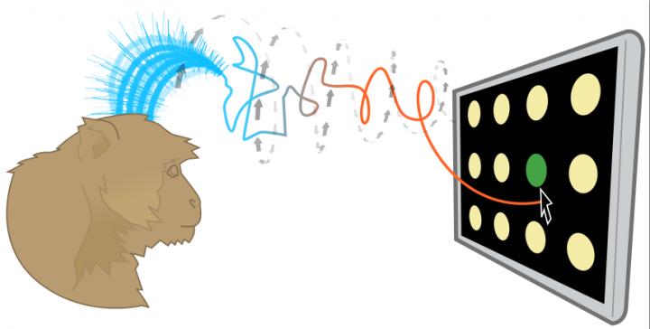
Brain-controlled prostheses sample a few hundred neurons to estimate motor commands that involve millions of neurons. So tiny sampling errors can reduce the precision and speed of thought-controlled keypads. A Stanford technique can analyze this sample and quickly make dozens of corrective adjustments to make thought control more precise. (credit: Jonathan Kao, Shenoy Lab)

An interdisciplinary team led by Stanford electrical engineer Krishna Shenoy has developed a technique to improve brain-controlled prostheses. These brain-computer-interface (BCI) devices, for people with neurological disease or spinal cord injury, deliver thought commands to devices such as virtual keypads, bypassing the damaged area.

The new technique addresses a problem with these brain-controlled prostheses: they currently access a sample of only a few hundred neurons, so tiny errors in the sample — neurons that fire too fast or too slow — reduce the precision and speed of thought-controlled keypads.

Understanding brain dynamics for arm movements

In essence the new prostheses analyze the neuron sample and quickly make dozens of corrective adjustments to the estimate of the brain’s electrical pattern.

Shenoy’s team tested a brain-controlled cursor meant to operate a virtual keyboard. The system is intended for people with paralysis and amyotrophic lateral sclerosis (ALS), also called Lou Gehrig’s disease, a condition that Stephen Hawking has. ALS degrades one’s ability to move.

The new corrective technique is based on a recently discovered understanding of how monkeys naturally perform arm movements. The researchers studied animals that were normal in every way. The monkeys used their arms, hands and fingers to reach for targets presented on a video screen. The researchers sought to learn, through hundreds of experiments, what the electrical patterns from the 100- to 200-neuron sample looked like during a normal reach — to understand the “brain dynamics” underlying reaching arm movements.

“These brain dynamics are analogous to rules that characterize the interactions of the millions of neurons that control motions,” said Jonathan Kao, a doctoral student in electrical engineering and first author of the open-access Nature Communications paper on the research. “They enable us to use a tiny sample more precisely.”

A decoding algorithm

In their current experiments, Shenoy’s team members distilled their understanding of brain dynamics into an algorithm that could decode (analyze) the measured electrical signals that their prosthetic device obtained from the sampled neurons. The algorithm tweaked these measured signals so that the sample’s dynamics were more like the baseline brain dynamics and thus more precise.

To test this algorithm, the Stanford researchers first trained two monkeys to choose targets on a simplified keypad. The keypad consisted of several rows and columns of blank circles. When a light flashed on a given circle the monkeys were trained to reach for that circle with their arms.

To set a performance baseline, the researchers measured how many targets the monkeys could tap with their fingers in 30 seconds. The monkeys averaged 29 correct finger taps in 30 seconds.

In the actual experiment, the researchers only scored virtual taps that came from the monkeys’ brain-controlled cursor. Although the monkey may still have moved his fingers, the researchers only counted a hit when the brain-controlled cursor, corrected by the algorithm, sent the virtual cursor to the target.

The prosthetic scored 26 thought-taps in 30 seconds, about 90 percent as quickly as a monkey’s finger. (See video of hand versus thought-controlled cursor taps.)

Thought-controlled keypads are not unique to Shenoy’s lab. Other brain-controlled prosthetics use different techniques to solve the problem of sampling error. But of several alternative techniques tested by the Stanford team, the closest resulted in 23 targets in 30 seconds.

Next steps

The goal of all this research is to get thought-controlled prosthetics to people with ALS. Today these people may use an eye-tracking system to direct cursors or a “head mouse” that tracks the movement of the head. Both are fatiguing to use. Neither provides the natural and intuitive control of readings taken directly from the brain.

“Brain-controlled prostheses will lead to a substantial improvement in quality of life,” Shenoy said. “The speed and accuracy demonstrated in this prosthesis results from years of basic neuroscience research and from combining these scientific discoveries with the principled design of mathematical control algorithms.”

The U.S. Food and Drug Administration recently gave Shenoy’s team the green light to conduct a pilot clinical trial of their thought-controlled cursor on people with spinal cord injuries.

“This is a fundamentally new approach that can be further refined and optimized to give brain-controlled prostheses greater performance, and therefore greater clinical viability,” Shenoy said.


Abstract of Single-trial dynamics of motor cortex and their applications to brain-machine interfaces

Increasing evidence suggests that neural population responses have their own internal drive, or dynamics, that describe how the neural population evolves through time. An important prediction of neural dynamical models is that previously observed neural activity is informative of noisy yet-to-be-observed activity on single-trials, and may thus have a denoising effect. To investigate this prediction, we built and characterized dynamical models of single-trial motor cortical activity. We find these models capture salient dynamical features of the neural population and are informative of future neural activity on single trials. To assess how neural dynamics may beneficially denoise single-trial neural activity, we incorporate neural dynamics into a brain–machine interface (BMI). In online experiments, we find that a neural dynamical BMI achieves substantially higher performance than its non-dynamical counterpart. These results provide evidence that neural dynamics beneficially inform the temporal evolution of neural activity on single trials and may directly impact the performance of BMIs.

Unlikely graphene-nanotube combination forms high-speed digital switch

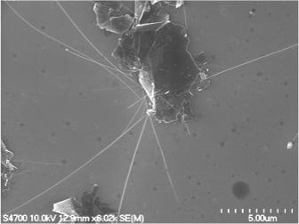
Hair-like boron nitride nanotubes intersect a sheet of graphene (top) to create a high-speed digital switch (credit: Michigan Tech, Yoke Khin Yap)

By themselves, graphene is too conductive while boron nitride nanotubes are too insulating, but combining them could create a workable digital switch — which can be used for controlling electrons in computers and other electronic devices.

To create this serendipitous super-hybrid, Yoke Khin Yap, a professor of physics at Michigan Technological University, and his team exfoliated (peeled off) graphene(from graphite) and modified the material’s surface with tiny pinholes, then grew the boron nitride nanotubes up and through the pinholes — like a plant randomly poking up through a crack in a concrete pavement. That formed a “band gap” mismatch, which created “a potential barrier  that stops electrons,” he said.

In other words, a switch.

The chemical structures of graphene (gray) and boron nitride nanotubes (pink and purple) can be used to create a digital switch at the point where the two materials come in contact (credit: Michigan Tech, Yoke Khin Yap)

High switching speed

The band gap mismatch results from the materials’ structure: graphene’s flat sheet conducts electricity quickly, and the atomic structure in the nanotubes halts electric currents. This disparity creates a barrier, caused by the difference in electron movement as currents move next to and past the hair-like boron nitride nanotubes. These points of contact between the materials, called heterojunctions, are what make the digital on/off switch possible.

Yap and his research team have also shown that because the materials are respectively so effective at conducting or stopping electricity, the resulting switching ratio is high. So how fast the materials can turn on and off is several orders of magnitude greater than current graphene switches. And this speed could eventually quicken the pace of electronics and computing.

Yap says this study is a continuation of past research into making transistors without semiconductors. The problem with semiconductors like silicon is that they can only get so small, and they give off a lot of heat; the use of graphene and nanotubes bypasses those problems. In addition, the graphene and boron nitride nanotubes have the same atomic arrangement pattern, or lattice matching. With their aligned atoms, the graphene-nanotube digital switches could avoid the issues of electron scattering.

“You want to control the direction of the electrons,” Yap explains, comparing the challenge to a pinball machine that traps, slows down and redirects electrons. “This is difficult in high speed environments, and the electron scattering reduces the number and speed of electrons.”

The journal Scientific Reports recently published their work in an open-access paper.


Abstract of Switching Behaviors of Graphene-Boron Nitride Nanotube Heterojunctions

High electron mobility of graphene has enabled their application in high-frequency analogue devices but their gapless nature has hindered their use in digital switches. In contrast, the structural analogous, h-BN sheets and BN nanotubes (BNNTs) are wide band gap insulators. Here we show that the growth of electrically insulating BNNTs on graphene can enable the use of graphene as effective digital switches. These graphene-BNNT heterojunctions were characterized at room temperature by four-probe scanning tunneling microscopy (4-probe STM) under real-time monitoring of scanning electron microscopy (SEM). A switching ratio as high as 105 at a turn-on voltage as low as 0.5 V were recorded. Simulation by density functional theory (DFT) suggests that mismatch of the density of states (DOS) is responsible for these novel switching behaviors.

Sleeping on your side may clear waste from your brain most effectively

The brain’s glymphatic pathway clears harmful wastes, especially during sleep. This lateral position could prove to be the best position for the brain-waste clearance process (credit: Stony Brook University)

Sleeping in the lateral, or side position, as compared to sleeping on one’s back or stomach, may more effectively remove brain waste, and could reduce the chances of developing Alzheimer’s, Parkinson’s and other neurological diseases, according to researchers at Stony Brook University.

Stony Brook University researchers discovered this in experiments with rodents by using dynamic contrast magnetic resonance imaging (MRI) to image the brain’s glymphatic pathway, a complex system that clears wastes and other harmful chemical solutes from the brain. They also used kinetic modeling to quantify the CSF-ISF exchange rates in anesthetized rodents’ brains in lateral, prone, and supine positions.

Colleagues at the University of Rochester used fluorescence microscopy and radioactive tracers to validate the MRI data and to assess the influence of body posture on the clearance of amyloid from the brains.

Their finding is published in the Journal of Neuroscience.

Most popular position in humans and animals

“It is interesting that the lateral sleep position is already the most popular in human and most animals —even in the wild — and it appears that we have adapted the lateral sleep position to most efficiently clear our brain of the metabolic waste products that built up while we are awake,” says Maiken Nedergaard, PhD, a co-author at the University of Rochester.

“The study therefore adds further support to the concept that sleep subserves a distinct biological function of sleep and that is to ‘clean up’ the mess that accumulates while we are awake. Many types of dementia are linked to sleep disturbances, including difficulties in falling asleep. It is increasing acknowledged that these sleep disturbances may accelerate memory loss in Alzheimer’s disease.”

The brain-waste clearing system

Cerebrospinal fluid (CSF) filters through the brain and exchanges with interstitial fluid (ISF) to clear waste in the glymphatic pathway, similar to the way the body’s lymphatic system clears waste from organs. The glymphatic pathway is most efficient during sleep. Brain waste includes amyloid β (amyloid) and tau proteins, chemicals that negatively affect brain processes if they build up.

Helene Benveniste, MD, PhD, Principal Investigator and a Professor in the Departments of Anesthesiology and Radiology at Stony Brook University School of Medicine, cautioned that further testing with MRI or other imaging methods in humans is necessary.

New York University Langone Medical Center was also involved in the research.


Abstract of The Effect of Body Posture on Brain Glymphatic Transport

The glymphatic pathway expedites clearance of waste, including soluble amyloidβ (Aβ) from the brain. Transport through this pathway is controlled by the brain’s arousal level because, during sleep or anesthesia, the brain’s interstitial space volume expands (compared with wakefulness), resulting in faster waste removal. Humans, as well as animals, exhibit different body postures during sleep, which may also affect waste removal. Therefore, not only the level of consciousness, but also body posture, might affect CSF–interstitial fluid (ISF) exchange efficiency. We used dynamic-contrast-enhanced MRI and kinetic modeling to quantify CSF-ISF exchange rates in anesthetized rodents” brains in supine, prone, or lateral positions. To validate the MRI data and to assess specifically the influence of body posture on clearance of Aβ, we used fluorescence microscopy and radioactive tracers, respectively. The analysis showed that glymphatic transport was most efficient in the lateral position compared with the supine or prone positions. In the prone position, in which the rat’s head was in the most upright position (mimicking posture during the awake state), transport was characterized by “retention” of the tracer, slower clearance, and more CSF efflux along larger caliber cervical vessels. The optical imaging and radiotracer studies confirmed that glymphatic transport and Aβ clearance were superior in the lateral and supine positions. We propose that the most popular sleep posture (lateral) has evolved to optimize waste removal during sleep and that posture must be considered in diagnostic imaging procedures developed in the future to assess CSF-ISF transport in humans.