Novel DNA origami structures

The versatility of the DNA origami 3D wireframe design technique created by Arizona State University Biodesign Institute researcher Hao Yan is demonstrated with the construction of this snub cube model, an Archimedean solid with 60 edges, 24 vertices and 38 faces including 6 squares and 32 equilateral triangles. (credit: TED-43/Wikimedia Commons)

Hao Yan, a researcher at Arizona State University’s Biodesign Institute, has extended DNA origami — which uses combinations of DNA base pairs to create new 2-D and 3-D nanoforms — into imaginative new forms that may one day lead to microelectronics and biomedical innovations.

“Earlier design methods [for DNA origami] used strategies including parallel arrangement of DNA helices to approximate arbitrary shapes, but precise fine-tuning of DNA wireframe architectures that connect vertices in 3D space has required a new approach,” says Yan, the Milton D. Glick Distinguished Chair of Chemistry and Biochemistry at ASU and directs Biodesign’s Center for Molecular Design and Biomimetics.

The new study describes wireframe structures of high complexity and programmability that are fabricated by precise control of branching and curvature, using novel organizational principles for the designs. (Wireframes are skeletal three-dimensional models represented purely through lines and vertices.)

The resulting nanoforms include symmetrical lattice arrays, quasicrystalline structures, curvilinear arrays, and a simple wire art sketch in the 100-nm scale, as well as 3-D objects including a snub cube with 60 edges and 24 vertices and a reconfigurable Archimedean solid that can be controlled to make the unfolding and refolding transitions between 3D and 2D.

The research appears in the advanced online edition of the journal Nature Nanotechnology.


DNA origami: how it works

In previous investigations, the Yan group created subtle architectural forms at an astonishingly minute scale, some measuring only tens of nanometers across — roughly the diameter of a virus particle. These nano-objects include spheres, spirals, flasksMöbius forms, and even an autonomous spider-like robot capable of following a prepared DNA track.
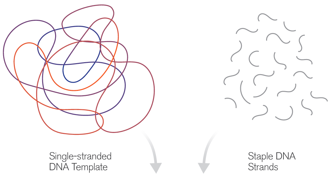
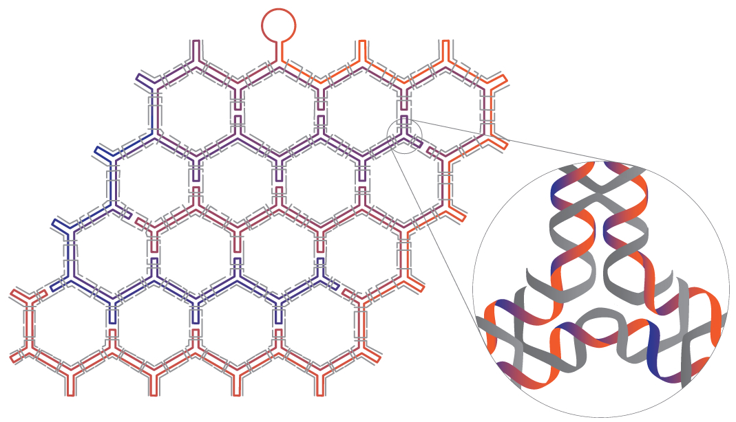
(credit: Fei Zhang et al./Nature Nanotechnology)

The technique of DNA origami capitalizes on the simple base-pairing properties of DNA, a molecule built from the four nucleotides Adenine (A), Thymine (T), Cytosine (C) and Guanine (G). The rules of the game are simple: A’s always pair with T’s and C’s with G’s. Using this abbreviated vocabulary, the myriad body plans of all living organisms are constructed; though duplicating even Nature’s simpler designs has required great ingenuity.

The basic idea of DNA origami is to use a length of single-stranded DNA as a scaffold for the desired shape. Base-pairing of complementary nucleotides causes the form to fold and self-assemble. The process is guided by the addition of shorter “staple strands,” which act to help fold the scaffold and to hold the resulting structure together. Various imaging technologies are used to observe the tiny structures, including fluorescence-, electron- and atomic force microscopy.


DNA origami originally produced nanoarchitectures of purely aesthetic interest, but refinements of the technique have opened the door to a new range of exciting applications including molecular cages for the encapsulation of molecules, enzyme immobilization and catalysis, chemical and biological sensing tools, drug delivery mechanisms, and molecular computing devices.

The technique described in the new study takes this approach a step further, allowing researchers to overcome local symmetry restrictions, creating wireframe architectures with higher order arbitrariness and complexity. Here, each line segment and vertex is individually designed and controlled. The number of arms emanating from each vertex may be varied from 2 to 10 and the precise angles between adjacent arms can be modified.
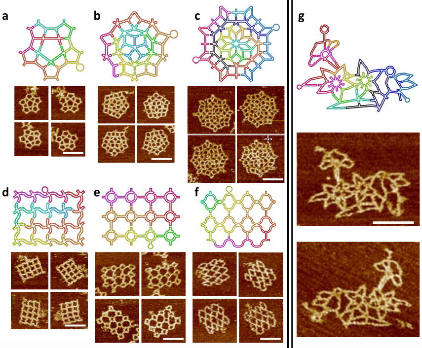
The scaffold folding path and representative AFM images for intricate 2D patterns. (a) A star-shape pattern without translational symmetry. (a) A Penrose tiling. (c) An 8-fold quasicrystalline pattern. (d-f) Three curved structures. (d) A waving grid. (e) A sphere array. (f) A fishnet. (g) A flower-and-bird pattern. All scale bars are 100 nm. Two scaffolds (one colorful and one black-blue) are used in c and g. (credit: Fei Zhang et al./Nature Nanotechnology)

In the current study, the method was first applied to symmetrical, regularly repeating polygonal designs, including hexagonal, square and triangular tiling geometries. Such common designs are known as tessellation patterns. A clever strategy involving a series of bridges and loops was used to properly route the scaffold strand, allowing it to pass through the entire structure, touching all lines of the wireframe once and only once. Staple strands were then applied to complete the designs.

In subsequent stages, the researchers created more complex wireframe structures, without the local translational symmetry found in the tessellation patterns. Three such patterns were made, including a star shape, a 5-fold Penrose tile and an 8-fold quasicrystalline pattern. (Quasicrystals are structures that are highly ordered but non-periodic. Such patterns can continuously fill available space, but are not translationally symmetric.)

Loop structures inserted into staple strands and unpaired nucleotides at the vertex points of the scaffold strands were also used, allowing researchers to perform precision modifications to the angles of junction arms.

The new design rules were next tested with the assembly of increasingly complex nanostructures, involving vertices ranging from 2 to 10 arms, with many different angles and curvatures involved, including a complex pattern of birds and flowers. The accuracy of the design was subsequently confirmed by AFM imaging, proving that the method could successfully yield highly sophisticated wireframe DNA nanostructures.

3D wire-frame Archimedean solid structures. (a) A 3D model of an Archimedean solid cuboctahedron with 12 vertices and 24 edges. Each vertex is a 4 Å~ 4 junction, and each edge is a 14-turn long double DNA duplex. (b) Left: Models showing possible conformations of the structure when deposited on mica surface; Right: the corresponding AFM images. (c) The reconfiguration between 3D and 2D can be realized by strand displacement by adding fuel and set strands. Top: reconfiguration schematics, Bottom: AFM images showing the transition. All scale bars in AFM images are 100 nm. (d) A 3D model of snub cube with 24 vertices and 60 edges. Each vertex is 5 Å~ 4 junction, and each edge is a 5-turn double DNA duplex. (e) Three views of the DNA snub cube from the design model (top of page). (credit: Fei Zhang et al./Nature Nanotechnology)

The method was then adapted to produce a number of 3D structures as well, including a cuboctahedron, and another Archimedian solid known as a snub cube — a structure with 60 edges, 24 vertices and 38 faces, including 6 squares and 32 equilateral triangles.

The authors stress that the new design innovations described can be used to compose and construct any imaginable wireframe nanostructure— a significant advancement for the burgeoning field.

On the horizon, nanoscale structures may one day be marshaled to hunt cancer cells in the body or act as robot assembly lines for the design of new drugs.


Abstract of Complex wireframe DNA origami nanostructures with multi-arm junction vertices

Structural DNA nanotechnology and the DNA origami technique, in particular, have provided a range of spatially addressable two- and three-dimensional nanostructures. These structures are, however, typically formed of tightly packed parallel helices. The development of wireframe structures should allow the creation of novel designs with unique functionalities, but engineering complex wireframe architectures with arbitrarily designed connections between selected vertices in three-dimensional space remains a challenge. Here, we report a design strategy for fabricating finite-size wireframe DNA nanostructures with high complexity and programmability. In our approach, the vertices are represented by n × 4 multi-arm junctions (n = 2–10) with controlled angles, and the lines are represented by antiparallel DNA crossover tiles of variable lengths. Scaffold strands are used to integrate the vertices and lines into fully assembled structures displaying intricate architectures. To demonstrate the versatility of the technique, a series of two-dimensional designs including quasi-crystalline patterns and curvilinear arrays or variable curvatures, and three-dimensional designs including a complex snub cube and a reconfigurable Archimedean solid were constructed.

An anti-inflammatory ‘smart drug’ that activates only in high-inflammation areas

Ben-Gurion University of the Negev (BGU) in Israel and University of Colorado researchers have developed a dynamic anti-inflammatory “smart” drug that can target specific sites in the body and could enhance the body’s natural ability to fight infection while reducing side effects.

This protein molecule, reported in the current issue of Journal of Immunology, has an exceptional property: when injected, it’s non-active. But upon reaching a local site with excessive inflammation, it becomes activated. Most other anti-inflammatory agents have broad effects in the body.

“This development is important because inhibition of inflammation in a non-specific manner reduces the natural ability to fight infections and is a common side effect of anti-inflammatory biologic therapeutics,” says Dr. Peleg Rider of BGU’s Department of Clinical Biochemistry and Pharmacology.

Using such a non-specific agent means any patient who suffers from local inflammation could be exposed to opportunistic infections at distant sites, such as lungs, which could risk tuberculosis, for example. This is especially a concern for immunosuppressed patients, as well as older patients and patients undergoing chemotherapy as part of an anti-cancer treatment course.

Mimicking a natural inflammatory process

The “chimeric IL-1Ra” protein combines an anti-inflammatory protein (right) with a peptide (left) that inactivates the IL-1Ra protein — except when the IL-1Ra portion encounters inflammatory enzymes, resulting in cleaving and releasing anti-inflammatory molecules (credit: Peleg Rider et al./Journal of Immunology)

“The beauty of this invention lies in the use of a known natural biological code,” Rider explains. “We mimicked a natural process that occurs during inflammation.”

Here’s how it works. The protein molecule (known as Chimeric IL-1Ra) is actually a chimera, combining two protein domains — both originating from the potent inflammatory IL-1 (interleukin family) protein (a group of 11 cytokines). (IL-1 normally plays a central role in regulating both immune and inflammatory responses to infections.)

The first part of this chimeric protein (IL-1beta) keeps a potent IL-1 natural infllammatory inhibitor (known as IL-1Ra) inactive. But when the IL-1Ra protein molecule encounters inflammatory enzymes, it springs into action, overriding IL-1beta, and is cleaved (split open), releasing powerful active molecules to reduce inflammation.

Rider, along with BGU’s Dr. Eli Lewis and Prof. Charles Dinarello of the University of Colorado, demonstrated their findings in a mouse model of local inflammation. They showed that leukocytes, which infiltrate inflammatory sites, indeed activate the chimeric protein, which in turn reduces local inflammation. The extent of activation of the protein correlated with the amount of inflammatory stimuli.

“Thus, a point that is highly relevant to clinical practice arises. Upon resolution of inflammation, the activation of the protein is also reduced and side effects are avoided,” Rider explains.

The new chimeric molecule was patented by BGN Technologies, BGU’s technology transfer company, and by the University of Colorado.

The research was supported by the Kamin program of Israel’s Ministry of Economy’s Chief Scientist’s Office.


Abstract of  IL-1 Receptor Antagonist Chimeric Protein: Context-Specific and Inflammation-Restricted Activation

Both IL-1α and IL-1β are highly inflammatory cytokines mediating a wide spectrum of diseases. A recombinant form of the naturally occurring IL-1R antagonist (IL-1Ra), which blocks IL-1R1, is broadly used to treat autoimmune and autoinflammatory diseases; however, blocking IL-1 increases the risk of infection. In this study, we describe the development of a novel form of recombinant IL-1Ra, termed chimeric IL-1Ra. This molecule is a fusion of the N-terminal peptide of IL-1β and IL-1Ra, resulting in inactive IL-1Ra. Because the IL-1β N-terminal peptide contains several protease sites clustered around the caspase-1 site, local proteases at sites of inflammation can cleave chimeric IL-1Ra and turn IL-1Ra active. We demonstrate that chimeric IL-1Ra reduces IL-1–mediated inflammation in vitro and in vivo. This unique approach limits IL-1 receptor blockade to sites of inflammation, while sparing a multitude of desired IL-1–related activities, including host defense against infections and IL-1–mediated repair.

NASA discovers first near-Earth-size planet in the habitable zone around a Sun-like star

This artist’s concept compares Earth (left) to the new planet, called Kepler-452b, which is about 60 percent larger in diameter (credits: NASA/JPL-Caltech/T. Pyle)

NASA’s Kepler mission has discovered the first near-Earth-size planet in the “habitable zone” around a Sun-like star. This discovery joins 11 other new small habitable zone candidate planets, marking another milestone in the journey to find another “Earth.”

The newly discovered Kepler-452b, located 1,400 light-years away in the constellation Cygnus, is the smallest planet to date discovered orbiting in the habitable zone — the area around a star where liquid water could pool on the surface of an orbiting planet — of a G2-type star, like our sun. The confirmation of Kepler-452b brings the total number of confirmed planets to 1,030.

“It’s awe-inspiring to consider that this planet has spent 6 billion years in the habitable zone of its star; longer than Earth,” said Jon Jenkins, Kepler data analysis lead at NASA’s Ames Research Center, who led the team that discovered Kepler-452b. ” That’s substantial opportunity for life to arise, should all the necessary ingredients and conditions for life exist on this planet.”

The CRISPR craze: genome editing technologies poised to revolutionize medicine and industry

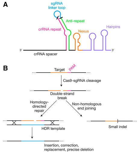
Genome editing by engineered Cas9 systems (credit: Mary Ann Liebert, Inc., publishers)

CRISPR/Cas systems for genome editing have revolutionized biological research over the past three years, and their ability to make targeted changes in DNA sequences in living cells with relative ease and affordability is now being applied to clinical medicine and will have a significant impact on advances in drug and other therapies, agriculture, and food products.

The power and promise of this innovation are presented in the Review article “The Bacterial Origins of the CRISPR Genome-Editing Revolution,” published in a special issue of Human Gene Therapy, a peer-reviewed journal from Mary Ann Liebert, Inc., publishers. The article is available open access until October 15, 2015 on the Human Gene Therapy website.

Erik Sontheimer, University of Massachusetts Medical School, Worcester, and Rodolphe Barrangou, North Carolina State University, Raleigh, describe the origins of this technology, which were derived from DNA sequences found in many bacteria known as clustered, regularly interspaced, short palindromic repeats (CRISPR) regions. These are part of bacteria’s protective immune system.

These regions have been developed into genome editing tools comprised of a “hardware” component (an RNA-guided DNA-targeting system that breaks a DNA strand at a specific site, with the help of the Cas protein), and a “software” component that can be programmed, and re-programmed, to repair or replace a faulty gene.

Impacts beyond academic research

“Although the CRISPR craze has yielded tremendous scientific progress and critical technological advances, it is important to keep in mind that the sgRNA–Cas9 technology is only 3 years old, and that notwithstanding current progress and momentum, we are yet to fully unleash the potential of these tools,” the authors note.

“Beyond academic research and the media, the most significant impact of CRISPR may well turn out to be in industry, with unprecedented levels of interest and investment from multiple distinct business segments, including pharmaceuticals and biotech, as well as covering the food supply chain from agriculture to livestock to other food products.”


Abstract of The Bacterial Origins of the CRISPR Genome-Editing Revolution

Like most of the tools that enable modern life science research, the recent genome-editing revolution has its biological roots in the world of bacteria and archaea. Clustered, regularly interspaced, short palindromic repeats (CRISPR) loci are found in the genomes of many bacteria and most archaea, and underlie an adaptive immune system that protects the host cell against invasive nucleic acids such as viral genomes. In recent years, engineered versions of these systems have enabled efficient DNA targeting in living cells from dozens of species (including humans and other eukaryotes), and the exploitation of the resulting endogenous DNA repair pathways has provided a route to fast, easy, and affordable genome editing. In only three years after RNA-guided DNA cleavage was first harnessed, the ability to edit genomes via simple, user-defined RNA sequences has already revolutionized nearly all areas of biological science. CRISPR-based technologies are now poised to similarly revolutionize many facets of clinical medicine, and even promise to advance the long-term goal of directly editing genomic sequences of patients with inherited disease. In this review, we describe the biological and mechanistic basis for these remarkable immune systems, and how their engineered derivatives are revolutionizing basic and clinical research.

Phosphorene could lead to ultrathin solar cells


Australian National University | Sticky tape the key to ultrathin solar cells

Scientists at Australian National University (ANU) have used simple transparent sticky (aka “Scotch”) tape to create single-atom-thick layers of phosphorene from “black phosphorus,” a black crystalline form of phosphorus similar to graphite (which is used to create graphene).

Unlike graphene, phosphorene is a natural semiconductor that can be switched on and off, like silicon, as KurzweilAI has reported. “Because phosphorene is so thin and light, it creates possibilities for making lots of interesting devices, such as LEDs or solar cells,” said lead researcher Yuerui (Larry) Lu, PhD, from ANU College of Engineering and Computer Science.

Properties that vary with layer thickness

Phosphorene is a thinner and lighter semiconductor than silicon, and it has unusual light emission properties that vary widely with the thickness of the layers, which enables more flexibility for manufacturing. “This property has never been reported before in any other material,” said Lu.

Schematic of the “puckered honeycomb” crystal structure of black phosphorus (credit: Vahid Tayari/McGill University)

“By changing the number of layers [peeled off] we can tightly control the band gap, which determines the material’s properties, such as the color of LED it would make.* “You can see quite clearly under the microscope the different colors of the sample, which tells you how many layers are there,” said Dr Lu.

The study was recently described in an open-access paper in the Nature journal Light: Science and Applications.

* Lu’s team found the optical gap for monolayer (single-layer) phosphorene was 1.75 electron volts, corresponding to red light of a wavelength of 700 nanometers. As more layers were added, the optical gap decreased. For instance, for five layers, the optical gap value was 0.8 electron volts, a infrared wavelength of 1550 nanometres. For very thick layers, the value was around 0.3 electron volts, a mid-infrared wavelength of around 3.5 microns.


Abstract of Optical tuning of exciton and trion emissions in monolayer phosphorene

Monolayer phosphorene provides a unique two-dimensional (2D) platform to investigate the fundamental dynamics of excitons and trions (charged excitons) in reduced dimensions. However, owing to its high instability, unambiguous identification of monolayer phosphorene has been elusive. Consequently, many important fundamental properties, such as exciton dynamics, remain underexplored. We report a rapid, noninvasive, and highly accurate approach based on optical interferometry to determine the layer number of phosphorene, and confirm the results with reliable photoluminescence measurements. Furthermore, we successfully probed the dynamics of excitons and trions in monolayer phosphorene by controlling the photo-carrier injection in a relatively low excitation power range. Based on our measured optical gap and the previously measured electronic energy gap, we determined the exciton binding energy to be ~0.3 eV for the monolayer phosphorene on SiO2/Si substrate, which agrees well with theoretical predictions. A huge trion binding energy of ~100 meV was first observed in monolayer phosphorene, which is around five times higher than that in transition metal dichalcogenide (TMD) monolayer semiconductor, such as MoS2. The carrier lifetime of exciton emission in monolayer phosphorene was measured to be ~220 ps, which is comparable to those in other 2D TMD semiconductors. Our results open new avenues for exploring fundamental phenomena and novel optoelectronic applications using monolayer phosphorene.

3D-printing basic electronic components

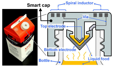
UC Berkeley engineers created a “smart cap” using 3-D-printed plastic with embedded electronics to wirelessly monitor the freshness of milk (credit: Photo and schematic by Sung-Yueh Wu)

UC Berkeley engineers, in collaboration with colleagues at Taiwan’s National Chiao Tung University, have developed a 3D printing process for creating basic electronic components, such as resistors, inductors, capacitors, and integrated wireless electrical sensing systems.

As a test, they printed a wireless “smart cap” for a milk carton that detected signs of spoilage using embedded sensors.

The findings were published Monday, July 20, in a new open-access journal in the Nature Publishing Group called Microsystems & Nanoengineering.

“Our paper describes the first demonstration of 3-D printing for working basic electrical components, as well as a working wireless sensor,” said senior author Liwei Lin, a professor of mechanical engineering and co-director of the Berkeley Sensor and Actuator Center.“One day, people may simply download 3D-printing files from the Internet with customized shapes and colors and print out useful devices at home.”

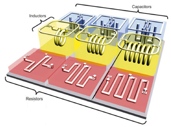
Engineers created a range of 3-D-printed electrical components, including an electrical resistor, inductor, capacitor and an LC tank (integrated inductor-capacitor system) (the penny is used for scale), shown here before removal of wax (credit: Photo by Sung-Yueh Wu)

The researchers started by printing polymers and wax. They then removed the wax, leaving hollow tubes into which liquid metal —- in their experiments they used silver — was injected and then cured.

The shape and design of the metal determined the function of different electrical components. For instance, thin wires acted as resistors, and flat plates were made into capacitors.

To demonstrate their use, the researchers integrated the electronic components into a plastic milk carton cap to monitor signs of spoilage. The “smart cap” was fitted with a capacitor and an inductor to form a resonant circuit. A quick flip of the carton allowed a bit of milk to get trapped in the cap’s capacitor gap, and the entire carton was then left unopened at room temperature (about 71.6 degrees Fahrenheit) for 36 hours.

3D-printed components (credit: Sung-Yueh Wu et al./Microsystems & Nanoengineering)

The circuit could detect the changes in electrical signals that accompany increased levels of bacteria. The researchers periodically monitored the changes with a wireless radio-frequency probe at the start of the experiment and every 12 hours thereafter, up to 36 hours. The property of milk changes gradually as it degrades, leading to variations in its electrical characteristics*.

Those changes were detected wirelessly using the smart cap, which found that the peak vibration frequency of the room-temperature milk dropped by 4.3 percent after 36 hours. In comparison, a carton of milk kept in refrigeration at 39.2 degrees Fahrenheit saw a relatively minor 0.12 percent shift in frequency over the same time period.

Cheap DIY electronic circuits 

“This 3D-printing technology could eventually make electronic circuits cheap enough to be added to packaging to provide food safety alerts for consumers,” said Lin. “You could imagine a scenario where you can use your cellphone to check the freshness of food while it’s still on the store shelves.”

As 3D printers become cheaper and better, the options for electronics will expand, said Lin, though he does not think people will be printing out their own smartphones or computers anytime soon.

“That would be very difficult because of the extremely small size of modern electronics,” he said. “It might also not be practical in terms of price since current integrated circuits are made by batch fabrication to keep costs low. Instead, I see 3D-printed microelectronic devices as very promising for systems that would benefit from customization.”

Lin said his lab is working on developing this technology for health applications, such as implantable devices with embedded transducers that can monitor blood pressure, muscle strain and drug concentrations.

* As the trapped milk hardened, the dielectric constant increased, increasing the capacitance and thus decreasing the frequency.


Abstract of 3D-printed microelectronics for integrated circuitry and passive wireless sensors

Three-dimensional (3D) additive manufacturing techniques have been utilized to make 3D electrical components, such as resistors, capacitors, and inductors, as well as circuits and passive wireless sensors. Using the fused deposition modeling technology and a multiple-nozzle system with a printing resolution of 30 μm, 3D structures with both supporting and sacrificial structures are constructed. After removing the sacrificial materials, suspensions with silver particles are injected subsequently solidified to form metallic elements/interconnects. The prototype results show good characteristics of fabricated 3D microelectronics components, including an inductor–capacitor-resonant tank circuitry with a resonance frequency at 0.53 GHz. A 3D “smart cap” with an embedded inductor–capacitor tank as the wireless passive sensor was demonstrated to monitor the quality of liquid food (e.g., milk and juice) wirelessly. The result shows a 4.3% resonance frequency shift from milk stored in the room temperature environment for 36 h. This work establishes an innovative approach to construct arbitrary 3D systems with embedded electrical structures as integrated circuitry for various applications, including the demonstrated passive wireless sensors.

Common chemicals may act together to increase cancer risk, international study finds

Disruptive potential of environmental exposures to mixtures of chemicals (credit: William H.Goodson III et al./Carcinogenesis)

Common environmental chemicals assumed to be safe at low doses may act separately or together to disrupt human tissues in ways that eventually lead to cancer, according to a task force of almost 200 scientists from 28 countries.

In a nearly three-year investigation of the state of knowledge about environmentally influenced cancers, the scientists studied low-dose effects of 85 common chemicals not considered to be carcinogenic to humans.

Common chemicals

The researchers reviewed the actions of these chemicals against a long list of mechanisms that are important for cancer development. Drawing on hundreds of laboratory studies, large databases of cancer information, and models that predict cancer development, they compared the chemicals’ biological activity patterns to 11 known cancer “hallmarks” – distinctive patterns of cellular and genetic disruption associated with early development of tumors.

The chemicals included bisphenol A (BPA), used in plastic food and beverage containers; rotenone, a broad-spectrum insecticide; paraquat, an agricultural herbicide; and triclosan, an antibacterial agent used in soaps and cosmetics.

In their survey, the researchers learned that 50 of the 85 chemicals had been shown to disrupt functioning of cells in ways that correlated with known early patterns of cancer, even at the low, presumably benign levels at which most people are exposed.

For 13 of them, the researchers found evidence of a dose-response threshold — a level of exposure at which a chemical is considered toxic by regulators. For 22, there was no toxicity information at all.

Synergistic effects over time

“Our findings also suggest these molecules may be acting in synergy to increase cancer activity,” said William Bisson, an assistant professor and cancer researcher at Oregon State University and a team leader on the study. For example, EDTA, a metal-ion-binding compound used in manufacturing and medicine, interferes with the body’s repair of damaged genes.

“EDTA doesn’t cause genetic mutations itself,” said Bisson, “but if you’re exposed to it along with some substance that is mutagenic, it enhances the effect because it disrupts DNA repair, a key layer of cancer defense.”

Bisson said the main purpose of this study was to highlight gaps in knowledge of environmentally influenced cancers and to set forth a research agenda for the next few years. He added that more research is still necessary to assess early exposure and to understand early stages of cancer development.

The study is part of the Halifax Project, sponsored by the Canadian nonprofit organization Getting to Know Cancer. The organization’s mission is to advance scientific knowledge about cancer linked to environmental exposures. The team’s findings are published in an open-access paper in a special issue of the journal Carcinogenesis.

Traditional risk assessment has historically focused on a quest for single chemicals and single modes of action — approaches that may underestimate cancer risk, said Bisson, an expert on computational chemical genomics (the modeling of biochemical molecular interactions in cancer processes). This study takes a different tack, examining the interplay over time of independent molecular processes triggered by low-dose exposures to chemicals.

“Cancer is a disease of diseases,” said Bisson. “It follows multi-step development patterns, and in most cases it has a long latency period. It has to be tackled from an angle that considers the complexity of these patterns.

“A better understanding of what’s driving things to the point where they get uncontrollable will be key for the development of effective strategies for prevention and early detection.”


Abstract of Assessing the carcinogenic potential of low-dose exposures to chemical mixtures in the environment: the challenge ahead

Lifestyle factors are responsible for a considerable portion of cancer incidence worldwide, but credible estimates from the World Health Organization and the International Agency for Research on Cancer (IARC) suggest that the fraction of cancers attributable to toxic environmental exposures is between 7% and 19%. To explore the hypothesis that low-dose exposures to mixtures of chemicals in the environment may be combining to contribute to environmental carcinogenesis, we reviewed 11 hallmark phenotypes of cancer, multiple priority target sites for disruption in each area and prototypical chemical disruptors for all targets, this included dose-response characterizations, evidence of low-dose effects and cross-hallmark effects for all targets and chemicals. In total, 85 examples of chemicals were reviewed for actions on key pathways/mechanisms related to carcinogenesis. Only 15% (13/85) were found to have evidence of a dose-response threshold, whereas 59% (50/85) exerted low-dose effects. No dose-response information was found for the remaining 26% (22/85). Our analysis suggests that the cumulative effects of individual (non-carcinogenic) chemicals acting on different pathways, and a variety of related systems, organs, tissues and cells could plausibly conspire to produce carcinogenic synergies. Additional basic research on carcinogenesis and research focused on low-dose effects of chemical mixtures needs to be rigorously pursued before the merits of this hypothesis can be further advanced. However, the structure of the World Health Organization International Programme on Chemical Safety ‘Mode of Action’ framework should be revisited as it has inherent weaknesses that are not fully aligned with our current understanding of cancer biology.

Deep Genomics launches, uniting deep learning and genome biology

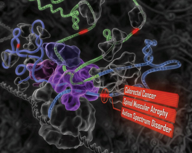
“Deep learning” reveals the genetic origins of disease. A computational system mimics the biology of RNA splicing by correlating DNA elements with splicing levels in healthy human tissues. The system can scan DNA and identify damaging genetic variants, including those deep within introns.This procedure has led to insights into the genetics of autism, cancers, and spinal muscular atrophy. (credit: Hui Y. Xiong et al./Science)

Deep Genomics, a University of Toronto spinoff, launched today (July 22), combining deep learning and artificial intelligence with the study of the human genome. The company is building on more than a decade of research and expertise in both fields.

Using deep learning allows Deep Genomics to predict the consequences of genomic alteration on various cell mechanisms to make life-changing decisions, potentially via personalized medicine treatment, the researchers say.

“Our vision is to change the course of genomic medicine and help save lives by determining smarter treatment options,” says Brendan Frey, the company’s president and CEO, a Fellow of the Canadian Institute for Advanced Research, and a Professor at the University of Toronto.

SPIDEX, a Google for DNA mutation effects

Professor Brendan Frey (center-right) and colleagues at the University of Toronto Faculty of Applied Science & Engineering (credit: Roberta Baker/ U of T Engineering)

The scientific community has spent decades searching for mutations within specific genes that can be connected to disease, such as the BRCA-1 and BRCA-2 genes for breast cancer. But there is a vast amount of mutations and combinations of mutations that have neither been observed nor studied, posing a challenge for diagnostics and therapeutics today.

“We envision a future where computers are trusted to predict the outcome of laboratory experiments and treatments, long before anyone picks up a test tube. Our first step will be to open up a genome-wide database of over 300 million potentially disease-causing variants, most of which are in regions of the genome that can’t be examined using other methods.”

Deep Genomics’ first product, called SPIDEX, provides information about how these DNA mutations may alter splicing in the cell, a process that is crucial for normal development. It also connects the dots between a variant or mutation of unknown significance and a variant that has been linked to a disease to determine its level of danger.

Because errant splicing is behind many diseases and disorders, including cancers and autism spectrum disorder, SPIDEX has immediate and practical importance for genetic testing and pharmaceutical development. The science validating the SPIDEX tool was described in the January 9, 2015 issue of the journal Science.

Labs will send the mutations they’ve collected to Deep Genomics, and the company will use their proprietary deep learning system, which includes SPIDEX, to “read” the genome and assess how likely the mutation is to cause a problem. SPIDEX can also connect the dots between a variant of unknown significance and a variant that has been linked to disease.

The company plans to further grow its team of machine learning, genome biology, and computational biology experts, and continue to invent new deep learning technologies and work with diagnosticians and biologists to understand the many complex ways that cells interpret DNA.

The company’s scientific advisory board includes Yann LeCun, Director, Facebook AI Research; Stephen Scherer, Director, The Center for Applied Genomics; and Jordan Lerner-Ellis, Director, Molecular Diagnostics at Mount Sinai Hospital.

More information: www.deepgenomics.com.


Abstract of The human splicing code reveals new insights into the genetic determinants of disease

To facilitate precision medicine and whole-genome annotation, we developed a machine-learning technique that scores how strongly genetic variants affect RNA splicing, whose alteration contributes to many diseases. Analysis of more than 650,000 intronic and exonic variants revealed widespread patterns of mutation-driven aberrant splicing. Intronic disease mutations that are more than 30 nucleotides from any splice site alter splicing nine times as often as common variants, and missense exonic disease mutations that have the least impact on protein function are five times as likely as others to alter splicing. We detected tens of thousands of disease-causing mutations, including those involved in cancers and spinal muscular atrophy. Examination of intronic and exonic variants found using whole-genome sequencing of individuals with autism revealed misspliced genes with neurodevelopmental phenotypes. Our approach provides evidence for causal variants and should enable new discoveries in precision medicine.

Korean researchers grow wafer-scale graphene on a silicon substrate

Wafer-scale (4 inch in diameter) synthesis of multi-layer graphene using high-temperature carbon ion implantation on nickel/SiO2/silicon (credit: J.Kim/Korea University, Korea)

Taking graphene a step closer to realistic commercial applications in silicon microelectronics, Korea University researchers have developed a simple microelectronics-compatible method for growing multi-layer graphene on a high-quality, wafer-scale (four inches in diameter) silicon substrate.

The method is based on the ion implantation technique — a process in which ions are accelerated under an electrical field and smashed into a semiconductor. The impacting ions change the physical, chemical, or electrical properties of the semiconductor.

Because of its high conductivity, “graphene is a potential contact electrode and an interconnection material linking semiconductor devices to form the desired electrical circuits, explained Jihyun Kim, the team leader and a professor in the Department of Chemical and Biological Engineering at Korea University.

However, “to deposit large-area graphene that is free of wrinkles, tears, and residues on silicon wafers requires low temperatures. That can’t be achieved with conventional chemical vapor deposition, which requires a high growth temperature — above 1,000 degrees Celsius.” That can cause strains, metal spiking, cracks, wrinkles, and contaminants from diffusion of dopants.

“Our synthesis method is controllable and scalable, allowing us to obtain graphene as large as the size of the silicon wafer,” Kim said. The researchers’ next step is to further lower the temperature in the synthesis process and to control the thickness of the graphene for manufacturing production.

The research is described in an open-access paper published this week in the journal Applied Physics Letters.


Abstract of Wafer-scale synthesis of multi-layer graphene by high-temperature carbon ion implantation

We report on the synthesis of wafer-scale (4 in. in diameter) high-quality multi-layer graphene using high-temperature carbon ion implantation on thin Ni films on a substrate of SiO2/Si.Carbon ions were bombarded at 20 keV and a dose of 1 × 1015 cm−2 onto the surface of the Ni/SiO2/Si substrate at a temperature of 500 °C. This was followed by high-temperature activation annealing (600–900 °C) to form a sp2-bonded honeycomb structure. The effects of post-implantation activation annealing conditions were systematically investigated by micro-Raman spectroscopy and transmission electron microscopy. Carbon ion implantation at elevated temperatures allowed a lower activation annealing temperature for fabricating large-area graphene. Our results indicate that carbon-ion implantation provides a facile and direct route for integrating graphene with Si microelectronics.

Deep neural network program recognizes sketches more accurately than a human

The Sketch-a-Net program successfully identified a seagull, pigeon, flying bird and standing bird better than humans (credit: QMUL, Mathias Eitz, James Hays and Marc Alexa)

The first computer program that can recognize hand-drawn sketches better than humans has been developed by researchers from Queen Mary University of London.

Known as Sketch-a-Net, the program correctly identified the subject of sketches 74.9 per cent of the time compared to humans that only managed a success rate of 73.1 per cent.

As sketching becomes more relevant with the increase in the use of touchscreens, it could lead to new ways to interact with computers. Touchscreens could understand what you are drawing enabling you to retrieve a specific image by drawing it with your fingers, which is more natural than keyword searches for finding items such as furniture or fashion accessories.

The improvement could also aid police forensics when an artist’s impression of a criminal needs to be matched to a mugshot or CCTV database.

The research also showed that the program performed better at determining finer details in sketches. For example, it was able to successfully distinguish “seagull,” “flying-bird,” “standing-bird” and “pigeon” with 42.5 per cent accuracy compared to humans, who only achieved 24.8 per cent.

Sketch-a-Net is a “deep neural network” program, designed to emulate the processing of the human brain. It is particularly successful because it accommodates the unique characteristics of sketches, particularly the order the strokes were drawn. This was information that was previously ignored but is especially important for understanding drawings on touchscreens.


Abstract of Sketch-a-Net that Beats Humans

We propose a multi-scale multi-channel deep neural network framework that, for the first time, yields sketch recognition performance surpassing that of humans. Our superior performance is a result of explicitly embedding the unique characteristics of sketches in our model: (i) a network architecture designed for sketch rather than natural photo statistics, (ii) a multi-channel generalisation that encodes sequential ordering in the sketching process, and (iii) a multi-scale network ensemble with joint Bayesian fusion that accounts for the different levels of abstraction exhibited in free-hand sketches. We show that state-of-the-art deep networks specifically engineered for photos of natural objects fail to perform well on sketch recognition, regardless whether they are trained using photo or sketch. Our network on the other hand not only delivers the best performance on the largest human sketch dataset to date, but also is small in size making efficient training possible using just CPUs.