Light electric stimulation of the brain may improve memory for people with schizophrenia

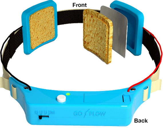
Transcranial direct-current stimulation device (credit: GoFlow)

Lightly stimulating the brain with transcranial direct current stimulation (tDCS) may improve short-term memory in people with schizophrenia, according to a new study by researchers at the Johns Hopkins University School of Medicine.

The tDCS procedure involves placing sponge-covered electrodes on the head and passing a weak electrical current between them.

David Schretlen, Ph.D., a professor of psychiatry and behavioral sciences at the Johns Hopkins University School of Medicine, reasoned that this type of brain stimulation might ease some of the cognitive difficulties that afflict people with schizophrenia.

A test based on prefrontal cortex stimulation

To test that possibility, Schretlen and Johns Hopkins colleagues targeted a brain region called the left dorsolateral prefrontal cortex, which plays an important role in short-term or working memory and is abnormal in people with schizophrenia, according to Schretlen.

Schretlen recruited 11 participants: five adults with confirmed schizophrenia and six of their close relatives (parents, siblings, and children of people with schizophrenia show some of the same abnormalities to a lesser degree, says Schretlen).

Each participant received two 30-minute treatments — one using a negative electrical charge, which the researchers thought might prove beneficial — and the other using a positive charge as a control. During and after each treatment, participants completed a battery of cognitive tests.

Thinking improvements

There were two notable results:

  • On tests of verbal and visual working memory, participants performed significantly better after receiving a negative charge, and the effects were “surprisingly strong,” says Schretlen.
  • Participants did better at the challenging task of switching between naming categories of items in a supermarket after a negatively charged treatment. The stimulation “was associated with better performance on working memory and subtle changes in word retrieval,” Schretlen says. People with schizophrenia often struggle to find the right words, he says. Because the prefrontal cortex contains a brain region responsible for word retrieval, Schretlen reasoned that transcranial direct current stimulation might help.

Schretlen is now studying transcranial direct current stimulation in a larger sample of patients using repeated sessions of stimulation, which he hopes will induce lasting benefits.

“Cognitive impairment is as ubiquitous as hallucinations in schizophrenia, yet medications only treat the hallucinations,” Schretlen says. “So even with medication, affected individuals often remain very disabled.” His hope is that transcranial direct current stimulation could give people with schizophrenia a shot at leading a more normal life.

Other findings

A related study last year showed that tDCS improved correction of mistakes. But another recent study found that after a repeated IQ test (which is normally expected to show improvements), IQ scores of people who underwent tDCS brain stimulation improved markedly less than did the IQ scores of people in the placebo group.

The tDCS procedure is also being studied by other researchers as a treatment for depression and Alzheimer’s-related memory loss, and to enhance recovery following strokes.

The research is described in a paper published online in Clinical Schizophrenia and Related Psychoses. The study was funded by the Therapeutic Cognitive Neuroscience Professorship; the Therapeutic Cognitive Neuroscience Fund; the Benjamin and Adith Miller Family Endowment on Aging, Alzheimer’s and Autism; and the National Institute on Child and Human Development.


Abstract of Can Transcranial Direct Current Stimulation Improve Cognitive Functioning in Adults with Schizophrenia?

Cognitive impairment is nearly ubiquitous in schizophrenia. First-degree relatives of persons with schizophrenia often show similar but milder deficits. Current methods for the treatment of schizophrenia are often ineffective in cognitive remediation. Since transcranial direct current stimulation (tDCS) can enhance cognitive functioning in healthy adults, it might provide a viable option to enhance cognition in schizophrenia. We sought to explore whether tDCS can be tolerated by persons with schizophrenia and potentially improve their cognitive functioning. We examined the effects of anodal versus cathodal tDCS on working memory and other cognitive tasks in five outpatients with schizophrenia and six first-degree relatives of persons with schizophrenia. Each participant completed tasks thought to be mediated by the prefrontal cortex during two 30-minute sessions of tDCS to the left and right dorsolateral prefrontal cortex (DLPFC). Anodal stimulation over the left DLPFC improved performance relative to cathodal stimulation on measures of working memory and aspects of verbal fluency relevant to word retrieval. The patient group showed differential changes in novel design production without alteration of overall productivity, suggesting that tDCS might be capable of altering selfmonitoring and executive control. All participants tolerated tDCS well. None withdrew from the study or experienced any adverse reaction. We conclude that adults with schizophrenia can tolerate tDCS while engaging in cognitive tasks and that tDCS can alter their performance.

A 99% biodegradable computer chip

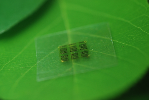
A cellulose nanofibril (CNF) computer chip shown on a leaf (credit: Yei Hwan Jung, Wisconsin Nano Engineering Device Laboratory)

University of Wisconsin-Madison and U.S. Department of Agriculture Forest Products Laboratory (FPL) researchers have jointly developed a wood chip in an effort to alleviate the environmental burden* of electronic devices.

Well, actually, a wood-substrate-based semiconductor chip. They replaced the silicon substrate portion in a conventional chip with environment-friendly cellulose nanofibril (CNF). CNF is a flexible, biodegradable material made from wood, as the researchers note in an open-access paper published May 26 in the journal Nature Communications.

“[More than 99%] of the material in a chip is support,” said Zhiyong Cai, project leader of an engineering composite science research group at FPL. With the new substrate, the chips are “so safe you can put them in the forest and fungus will degrade it. They become as safe as fertilizer.”

The new material is especially important for microwave chips (such as those used in mobile phones) made with gallium arsenide, which is especially difficult to fabricate on foreign substrates. That’s because of the small feature sizes and high temperature processes required for high performance.

Cai’s group addressed two key barriers to using wood-derived materials in an electronics setting: surface roughness and thermal expansion. “You don’t want it to expand or shrink too much. Wood is a natural hydroscopic [water-absorbing] material and could attract moisture from the air and expand,” Cai says. “With an epoxy coating on the surface of the CNF, we solved both [problems].”

* In 2007, it was estimated that over 426,000 cell phones (most of them were still functional) and 112,000 computers were discarded every day in the US, totalling 3.2 million tons of electronic waste generated per year, the researcher note in the paper.


Abstract of High-performance green flexible electronics based on biodegradable cellulose nanofibril paper

Today’s consumer electronics, such as cell phones, tablets and other portable electronic devices, are typically made of non-renewable, non-biodegradable, and sometimes potentially toxic (for example, gallium arsenide) materials. These consumer electronics are frequently upgraded or discarded, leading to serious environmental contamination. Thus, electronic systems consisting of renewable and biodegradable materials and minimal amount of potentially toxic materials are desirable. Here we report high-performance flexible microwave and digital electronics that consume the smallest amount of potentially toxic materials on biobased, biodegradable and flexible cellulose nanofibril papers. Furthermore, we demonstrate gallium arsenide microwave devices, the consumer wireless workhorse, in a transferrable thin-film form. Successful fabrication of key electrical components on the flexible cellulose nanofibril paper with comparable performance to their rigid counterparts and clear demonstration of fungal biodegradation of the cellulose-nanofibril-based electronics suggest that it is feasible to fabricate high-performance flexible electronics using ecofriendly materials.

Psychedelic drugs should be legally reclassified, says psychiatrist

The Persistence of Memory (credit: Salvador Dali)

Psychedelic drugs such as LSD are much less harmful than claimed and should be legally reclassified to allow further research on their medical use, says James Rucker, a psychiatrist and honorary lecturer at the Institute of Psychiatry, Psychology and Neuroscience, King’s College London.

These substances “were extensively used and researched in clinical psychiatry” before their prohibition in 1967 and many trials of these drugs in the 1950s and 1960s suggested “beneficial change in many psychiatric disorders.”

Nonetheless, in the UK, psychedelic drugs were legally classified as schedule 1 class A drugs; that is, as having “no accepted medical use and the greatest potential for harm, despite the research evidence to the contrary,” he writes.

Clinical efficacy shown in anxiety, obsessive compulsive disorder, addiction, and headaches

Rucker makes these points:

  • Psychedelics remain more legally restricted than heroin and cocaine. “But no evidence indicates that psychedelic drugs are habit forming; little evidence indicates that they are harmful in controlled settings; and much historical evidence shows that they could have use in common psychiatric disorders.” In fact, recent studies indicate that psychedelics have “clinical efficacy in anxiety associated with advanced cancer, obsessive compulsive disorder, tobacco and alcohol addiction, and cluster headaches,” he writes.
  • At present, larger clinical studies on psychedelics are made “almost impossible by the practical, financial and bureaucratic obstacles” imposed by their schedule 1 classification. Currently, only one manufacturer in the world produces psilocybin for trial purposes, he says, at a “prohibitive” cost of £100,000 for 1 g (50 doses).
  • In the UK, to hold a schedule 1 drug, institutions require a license, which costs about £5,000, he adds. Only four hospitals currently hold such licenses, which come with regular police or home office inspections and onerous rules on storage and transport, so “clinical research using psychedelics costs 5–10 times that of research into less restricted (but more harmful) drugs such as heroin.” As a result, “almost all grant funders are uncomfortable funding research into psychedelics,” while prohibition as a condition of UN membership is “arguably causing more harm than it prevents.”
  • Psychedelics are neither harmful nor addictive compared with other controlled substances. He calls on the UK Advisory Council on the Misuse of Drugs and the 2016 UN General Assembly Special Session on Drugs, “to recommend that psychedelics be reclassified as schedule 2 compounds to enable a comprehensive, evidence based assessment of their therapeutic potential.”


The Beatles — A Day in the Life

Medical ‘millirobots’ could replace invasive surgery

Cross-section: three-component Gauss gun before (top) and after (bottom) firing (credit: Aaron T. Becker et al./Proceedings of the IEEE)

University of Houston researchers have developed a concept for MRI-powered millimeter-size “millirobots” that could one day perform unprecedented minimally invasive medical treatments.

This technology could be used to treat hydrocephalus, for example. Current treatments require drilling through the skull to implant pressure-relieving shunts, said Aaron T. Becker, assistant professor of electrical and computer engineering at the University of Houston.

But MRI scanners alone don’t produce enough force to pierce tissues (or insert needles). So the researchers drew upon the principle of the “Gauss gun.”


K&J Magnetics | Gauss Gun Demonstrations

Here’s how the a Gauss gun works: a single steel ball rolls down a chamber, setting off a chain reaction when it smashes into the next ball, etc., until the last ball flies forward, moving much more quickly the initial ball.

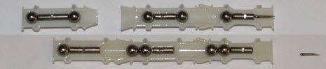
Based on that concept, the researchers imagine a medical robot with a barrel self-assembled from three small high-impact 3D-printed plastic components, with slender titanium rod spacers separating two steel balls.

Millirobot components (credit: Aaron T. Becker et al./Proceedings of the IEEE)

Aaron T. Becker, assistant professor of electrical and computer engineering at the University of Houston, said the potential technology could be used to treat hydrocephalus and other conditions, allowing surgeons to avoid current treatments that require cutting through the skull to implant pressure-relieving shunts.

Becker was first author of a paper presented at ICRA, the conference of the IEEE Robotics and Automation Society, nominated for best conference paper and best medical robotics paper.

“Hydrocephalus, among other conditions, is a candidate for correction by our millirobots because the ventricles are fluid-filled and connect to the spinal canal,” Becker said. “Our noninvasive approach would eventually require simply a hypodermic needle or lumbar puncture to introduce the components into the spinal canal, and the components could be steered out of the body afterwards.”

Future work will focus on exploring clinical context, miniaturizing the device, and optimizing material selection.


Abstract of Toward Tissue Penetration by MRI-powered Millirobots Using a Self-Assembled Gauss Gun

MRI-based navigation and propulsion of millirobots is a new and promising approach for minimally invasive therapies. The strong central field inside the scanner, however, precludes torque-based control. Consequently, prior propulsion techniques have been limited to gradient-based pulling through fluid-filled body lumens. This paper introduces a technique for generating large impulsive forces that can be used to penetrate tissue. The approach is based on navigating multiple robots to a desired location and using self-assembly to trigger the conversion of magnetic potential energy into sufficient kinetic energy to achieve penetration. The approach is illustrated through analytical modeling and experiments in a clinical MRI scanner.

New tech keeps your smart phone charged for 30 percent longer

(credit: iStock)

Engineers  at The Ohio State University claim they have created a circuit that makes cell phone batteries last up to 30 percent longer on a single charge. The trick: it converts some of the radio signals emanating from a phone into direct current (DC) power, which then charges the phone’s battery, they state.

This new technology can be built into a cell phone case, adding minimal bulk and weight.

“When we communicate with a cell tower or Wi-Fi router, so much energy goes to waste,” explained Chi-Chih Chen, research associate professor of electrical and computer engineering. “We recycle some of that wasted energy back into the battery.”

“Our technology is based on harvesting energy directly from the source, explained Robert Lee, professor of electrical and computer engineering. By Lee’s reckoning, nearly 97 percent of cell phone signals never reach a destination and are simply lost. Some of the that energy can be captured.

The idea is to siphon off just enough of the radio signal to noticeably slow battery drain, but not enough to degrade voice quality or data transmission. Cell phones broadcast in all directions at once to reach the nearest cell tower or Wi-Fi router. Chen and his colleagues came up with a system that identifies which radio signals are being wasted. It works only when a phone is transmitting.

Next, the engineers want to insert the device into a “skin” that sticks directly to a phone, or better, partner with a manufacturer to build it directly into a phone, tablet or other portable electronic device.

UPDATE June 6: Responding to a request for more information on energy harvesting, we received the following statement from Will Zell, CEO of licensee Nikola Labs: “Nikola Labs has a limit to the technical details we are able to share until our patents are published.”

Disney researchers develop 2-legged robot that walks like an animated character

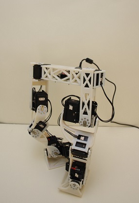
Robot mimics character’s movements (credit: Disney Research)

Disney researchers have found a way for a robot to mimic an animated character’s walk, bringing a cartoon (or other) character to life in the real world.

Beginning with an animation of a diminutive, peanut-shaped character that walks with a rolling, somewhat bow-legged gait, Katsu Yamane and his team at Disney Research Pittsburgh analyzed the character’s motion to design a robotic frame that could duplicate the walking motion. using 3D-printed links and servo motors, while also fitting inside the character’s skin. They then created control software that could keep the robot balanced while duplicating the character’s gait as closely as possible.

“The biggest challenge is that designers don’t necessarily consider physics when they create an animated character,” said Yamane, senior research scientist. Roboticists, however, wrestle with physical constraints throughout the process of creating a real-life version of the character.

“It’s important that, despite physical limitations, we do not sacrifice style or the quality of motion,” Yamane said. The robots will need to not only look like the characters, but move in the way people are accustomed to seeing those characters move.

(credit: Disney Research)

The researchers are describing the techniques and technologies they used to create the bipedal robot at the IEEE International Conference on Robotics and Automation, ICRA 2015, May 26–30 in Seattle.


DisneyResearchHub | Development of a Bipedal Robot that Walks Like an Animation Character

Intelligent handheld robots could make is easier for people to learn new skills

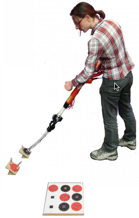
An intelligent handheld robot assisting a user in placing correct colored tiles (credit: University of Bristol)

What if your handheld tools knew what needs to be done and were even able to guide and help you complete jobs that require skills? University of Bristol researchers are finding out by building and testing intelligent handheld robots.

Think of them as smart power tools that “know” what they’re doing — and could even help you use them.

The robot tools would have three levels of autonomy, said Walterio Mayol-Cuevas, Reader in Robotics Computer Vision and Mobile Systems: “No autonomy, semi-autonomous — the robot advises the user but does not act, and fully autonomous — the robot advises and acts even by correcting or refusing to perform incorrect user actions.”

The Bristol team has experimented with tasks such as picking and dropping different objects to form tile patterns and aiming in 3D for simulated painting.

The robot designs are open source and available on the university’s HandheldRobotics page.


HandheldRobotics | The Design and Evaluation of a Cooperative Handheld Robot

A chip implanted under the skin allows for precise, real-time medical monitoring

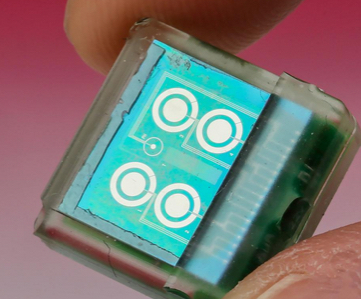
Under-the-skin chip (credit: EPFL)

A tiny (one-centimeter-square) biosensor chip developed at EPFL is designed to be implanted under your skin to continuously monitor concentrations of pH, temperature, and metabolism-related molecules like glucose, lactate and cholesterol, as well as some drugs.

The chip would replace blood work, which may take  hours — or even days — for analysis and is a limited snapshot of conditions at the moment the blood is drawn.

Developer Sandro Carrara unveiled the chip Tuesday (May 26) at the International Symposium on Circuits and Systems (ISCAS) in Lisbon.

The electrochemical sensors work with or without enzymes, which means the device can react to a wide range of compounds, and it can do so for several days or even weeks.

Wireless power and monitoring

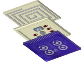
Implantable biosensor chip with three layers: a passive sensing platform (bottom), integrated circuits (middle) to analyze electrochemical measurements and generate a Bluetooth signal, and a coil (top) for through-the-skin data transmission and power via an external battery (credit: Camilla Baj-Rossi et al./IEEE Transactions on Biomedical Circuits and Systems)

The biochip contains three main components: a circuit with six sensors, a control unit that analyzes incoming signals, and a Bluetooth module for sending the results immediately to a mobile phone.

It also has an induction coil that wirelessly draws power from an external battery attached to the skin by a patch.

To ensure biocompatibility, an epoxy-enhanced polyurethane membrane was used to cover the device.

The chip was successfully tested in vivo on mice at the Institute for Research in Biomedicine (IRB) in Bellinzona, where researchers were able to constantly monitor glucose and paracetamol levels without a wire tracker getting in the way of the animals’ daily activities.

The results were promising, so clinical tests on humans could take place in three to five years — especially since the procedure is minimally invasive, the researchers say.

“Knowing the precise and real-time effect of drugs on the metabolism is one of the keys to the type of personalised, precision medicine that we are striving for,” said Carrara.

Dynamically reprogramming matter

Various types of reprogramming DNA strands can be used to selectively trigger transformations to radically different phases (configurations) of the initial particle structure (credit: Brookhaven National Laboratory)

Scientists at the U.S. Department of Energy’s Brookhaven National Laboratory have developed the capability of creating dynamic nanomaterials — ones whose structure and associated properties can be switched, on-demand. In a paper appearing in Nature Materials, they describe a way to selectively rearrange nanoparticles in three-dimensional arrays to produce different configurations, or “phases,” from the same nano-components.

“One of the goals in nanoparticle self-assembly has been to create structures by design,” said Oleg Gang, who led the work at Brookhaven’s Center for Functional Nanomaterials (CFN), a DOE Office of Science User Facility. “Until now, most of the structures we’ve built have been static.” KurzweilAI covered that development in a previous article, “Creating complex structures using DNA origami and nanoparticles.”

The new advance in nanoscale engineering builds on that previous work in developing ways to get nanoparticles to self-assemble into complex composite arrays, including linking them together with tethers constructed of complementary strands of synthetic DNA.

“We know that properties of materials built from nanoparticles are strongly dependent on their arrangements,” said Gang. “Previously, we’ve even been able to manipulate optical properties by shortening or lengthening the DNA tethers. But that approach does not permit us to achieve a global reorganization of the entire structure once it’s already built.”

DNA-directed rearrangement

“Now we are trying to achieve an even more ambitious goal,” reveal Gang: “Making materials that can transform so we can take advantage of properties that emerge with the particles’ rearrangements.”

The ability to direct particle rearrangements, or phase changes, will allow the scientists to choose the desired properties — say, the material’s response to light or a magnetic field — and switch them whenever needed. Such phase-changing materials could lead to radical new applications, such as dynamic energy-harvesting or responsive optical materials.

Injecting different kinds of reprogramming DNA strands can change the interparticle interactions in different ways depending on whether the new strands increase attraction or repulsion, or there’s a combination of these forces between particles (credit: Brookhaven National Laboratory)

In the new approach, the reprogramming DNA strands adhere to open binding sites on the already assembled nanoparticles. These strands exert additional forces on the linked-up nanoparticles.

“By introducing different types of reprogramming DNA strands, we modify the DNA shells surrounding the nanoparticles,” explained CFN postdoctoral fellow Yugang Zhang, the lead author on the paper. “Altering these shells can selectively shift the particle-particle interactions, either by increasing both attraction and repulsion, or by separately increasing only attraction or only repulsion. These reprogrammed interactions impose new constraints on the particles, forcing them to achieve a new structural organization to satisfy those constraints.”

Using their method, the team demonstrated that they could switch their original nanoparticle array, the “mother” phase, into multiple different daughter phases with precision control.

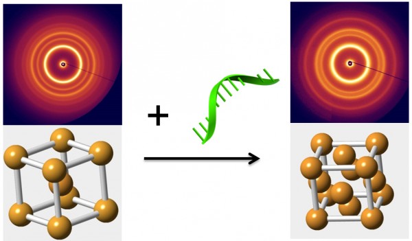
Introducing “reprogramming” of DNA strands in an already assembled nanoparticle array triggers a transition from a “mother phase,” where particles occupy the corners and center of a cube (left), to a more compact “daughter phase” (right). The change represented in the schematic diagrams is revealed by the associated small-angle x-ray scattering patterns. Such phase-changes could potentially be used to switch a material’s properties on demand. (credit: Brookhaven National Laboratory)

DNA-based matter reprogramming

This is quite different from phase changes driven by external physical conditions such as pressure or temperature, Gang said, which typically result in single phase shifts, or sometimes sequential ones. “In those cases, to go from phase A to phase C, you first have to shift from A to B and then B to C,” said Gang. “Our method allows us to pick which daughter phase we want and go right to that one because the daughter phase is completely determined by the type of DNA reprogramming strands we use.”

The scientists were able to observe the structural transformations to various daughter phases using a technique called in situ small-angle x-ray scattering at the National Synchrotron Light Source, a DOE Office of Science User Facility that operated at Brookhaven Lab from 1982 until last September (now replaced by NSLS-II, which produces x-ray beams 10,000 times brighter). The team also used computational modeling to calculate how different kinds of reprogramming strands would alter the interparticle interactions, and found their calculations agreed well with their experimental observations.

“The ability to dynamically switch the phase of an entire superlattice array will allow the creation of reprogrammable and switchable materials wherein multiple, different functions can be activated on demand,” said Gang. “Our experimental work and accompanying theoretical analysis confirm that reprogramming DNA-mediated interactions among nanoparticles is a viable way to achieve this goal.”

This research was done in collaboration with scientists from Columbia University’s School of Engineering and Applied Science and the Indian Institute of Technology Gandhinagar. The work was funded by the DOE Office of Science.


Abstract of Selective transformations between nanoparticle superlattices via the reprogramming of DNA-mediated interactions

The rapid development of self-assembly approaches has enabled the creation of materials with desired organization of nanoscale components. However, achieving dynamic control, wherein the system can be transformed on demand into multiple entirely different states, is typically absent in atomic and molecular systems and has remained elusive in designed nanoparticle systems. Here, we demonstrate with in situ small-angle X-ray scattering that, by using DNA strands as inputs, the structure of a three-dimensional lattice of DNA-coated nanoparticles can be switched from an initial ‘mother’ phase into one of multiple ‘daughter’ phases. The introduction of different types of reprogramming DNA strands modifies the DNA shells of the nanoparticles within the superlattice, thereby shifting interparticle interactions to drive the transformation into a particular daughter phase. Moreover, we mapped quantitatively with free-energy calculations the selective reprogramming of interactions onto the observed daughter phases.

Creating complex structures using DNA origami and nanoparticles

Cluster assembled from DNA-functionalized gold nanoparticles on vertices of a octahedral DNA origami frame (credit: Brookhaven National Laboratory))

Scientists at the U.S. Department of Energy’s (DOE) Brookhaven National Laboratory and collaborators have developed a method using DNA for designing new customized materials with complex structures for applications in energy, optics, and medicine.

They used ropelike configurations of DNA to form a rigid geometrical framework and then added dangling pieces of single-stranded DNA to glue nanoparticles in place.

The method, described in the journal Nature Nanotechnology, produced predictable geometric configurations that are somewhat analogous to molecules made of atoms, according to Brookhaven physicist Oleg Gang, who led the project at the Lab’s Center for Functional Nanomaterials (CFN).

“While atoms form molecules based on the nature of their chemical bonds, there has been no easy way to impose such a specific spatial binding scheme on nanoparticles, he said. “This is exactly the problem that our method addresses.

“We may be able to design materials that mimic nature’s machinery to harvest solar energy, or manipulate light for telecommunications applications, or design novel catalysts for speeding up a variety of chemical reactions,” Gang said.

As a demonstration, the researchers used an octahedral (eight-sided) scaffold (structure) with particles positioned in precise locations on the scaffold according to specific DNA coding. They also used the geometrical clusters as building blocks for larger arrays, including linear chains and two-dimensional planar sheets.

“Our work demonstrates the versatility of this approach and opens up numerous exciting opportunities for high-yield precision assembly of tailored 3D building blocks in which multiple nanoparticles of different structures and functions can be integrated,” said CFN scientist Ye Tian, one of the lead authors on the paper.

A new DNA “origami” kit

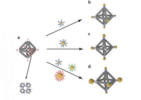
Scientists built octahedrons using ropelike structures made of bundles of DNA double-helix molecules to form the frames (a). Single strands of DNA attached at the vertices (numbered in red) can be used to attach nanoparticles coated with complementary strands. This approach can yield a variety of structures, including ones with the same type of particle at each vertex (b), arrangements with particles placed only on certain vertices (c), and structures with different particles placed strategically on different vertices (d). (credit: Brookhaven National Laboratory)

This nanoscale construction approach takes advantage of two key characteristics of the DNA molecule: the twisted-ladder double helix shape, and the natural tendency of strands with complementary bases (the A, T, G, and C letters of the genetic code) to pair up in a precise way.

Here’s how the scientists built a complex structure with this “DNA origami” kit:

1. They created bundles of six double-helix DNA molecules.

2. They put four of these bundles together to make a stable, somewhat rigid building material — similar to the way individual fibrous strands are woven together to make a very strong rope.

3. They used these ropelike girders to form the frame of three-dimensional octahedrons, “stapling” the linear DNA chains together with hundreds of short complementary DNA strands. (“We refer to these as DNA origami octahedrons,” Gang said.)

4. To make it possible to “glue” nanoparticles to the 3D frames, the scientists engineered each of the original six-helix bundles to have one helix with an extra single-stranded piece of DNA sticking out from both ends.

5. When assembled into the 3D octahedrons, each vertex of the frame had a few of these “sticky end” tethers available for binding with objects coated with complementary DNA strands.

“When nanoparticles coated with single strand tethers are mixed with the DNA origami octahedrons, the ‘free’ pieces of DNA find one another so the bases can pair up according to the rules of the DNA complementarity code. Thus the specifically DNA-encoded particles can find their correspondingly designed place on the octahedron vertices,” Gang explained.

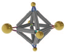
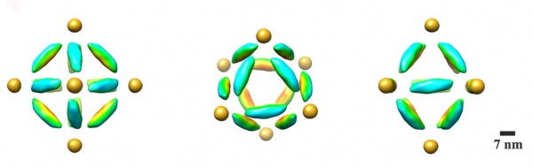
A combination cryo-electron microscopy image of an octahedral frame with one gold nanoparticle bound to each of the six vertices, shown from three different angles. (Credit: Brookhaven National Laboratory)

The scientists can also change what binds to each vertex by changing the DNA sequences encoded on the tethers. In one experiment, they encoded the same sequence on all the octahedron’s tethers, and attached strands with a complementary sequence to gold nanoparticles. The result: One gold nanoparticle attached to each of octahedron’s six vertices.

In additional experiments,the scientists changed the sequence of some vertices and used complementary strands on different kinds of particles, illustrating that they could direct the assembly and arrangement of the particles in a very precise way.

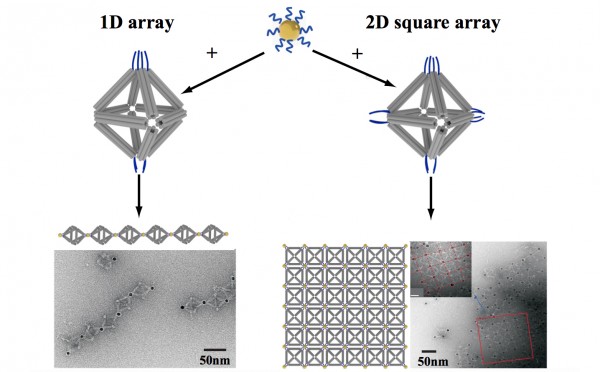
By strategically placing tethers on particular vertices, the scientists used the octahedrons to link nanoparticles into one-dimensional chainlike arrays (left) and two-dimensional square sheets (right). (Credit: Brookhaven National Laboratory)

In one case, they made two different arrangements of the same three pairs of particles of different sizes, producing products with different optical properties. They were even able to use DNA tethers on selected vertices to link octahedrons end-to-end, forming chains, and in 2D arrays, forming sheets.

Visualizing the structures

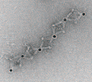
TEM image of part of the 1D array (credit: Brookhaven National Lab)

Confirming the particle arrangements and structures was a major challenge because the nanoparticles and the DNA molecules making up the frames have very different densities. Certain microscopy techniques would reveal only the particles, while others would distort the 3D structures.

To see both the particles and origami frames, the scientists used cryo-electron microscopy (cryo-EM), led by Brookhaven Lab and Stony Brook University biologist Huilin Li, an expert in this technique, and Tong Wang, the paper’s other lead co-author, who works in Brookhaven’s Biosciences department with Li.

They had to subtract information from the images to “see” the different density components separately, then combine the information using single particle 3D reconstruction and tomography to produce the final images.

This research was supported by the DOE Office of Science.


Abstract of Prescribed nanoparticle cluster architectures and low-dimensional arrays built using octahedral DNA origami frames

Three-dimensional mesoscale clusters that are formed from nanoparticles spatially arranged in pre-determined positions
can be thought of as mesoscale analogues of molecules. These nanoparticle architectures could offer tailored properties
due to collective effects, but developing a general platform for fabricating such clusters is a significant challenge. Here, we
report a strategy for assembling three-dimensional nanoparticle clusters that uses a molecular frame designed with
encoded vertices for particle placement. The frame is a DNA origami octahedron and can be used to fabricate clusters
with various symmetries and particle compositions. Cryo-electron microscopy is used to uncover the structure of the DNA
frame and to reveal that the nanoparticles are spatially coordinated in the prescribed manner. We show that the DNA
frame and one set of nanoparticles can be used to create nanoclusters with different chiroptical activities. We also show
that the octahedra can serve as programmable interparticle linkers, allowing one- and two-dimensional arrays to be
assembled with designed particle arrangements.