Seeing the invisible: visible-light metamaterial superlens made from nanobeads

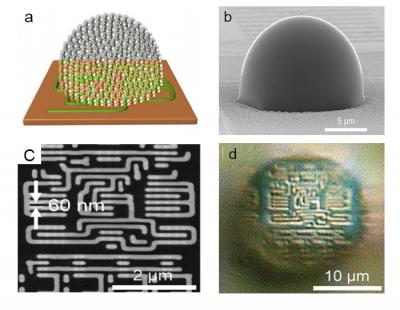
(a) Conceptual drawing of nanoparticle-based metamaterial solid immersion lens (mSIL) (b) Lab-made mSIL using titanium dioxide nanoparticles (c) SEM image of 60 nm size imaging sample (d) corresponding superlens imaging of the 60 nm sample by the developed mSIL. (credit: BangorUniversity/Fudan University)

A team of British and Chinese scientists has developed a new “metamaterial-based solid immersion lens” (mSIL) microscope lens design that can extend the magnification of an optical microscope to see objects smaller than the approximately 200 nanometers Abbe diffraction limit, the smallest size of bacteria.

Led by Zengbo Wang, PhD, at Bangor University UK and Prof Limin Wu at Fudan University, China, the team created minute droplet-like lens structures on the surface to be examined. These act as an additional lens to magnify the surface features previously invisible to a normal microscope lens, adding 5x magnification to existing microscopes.

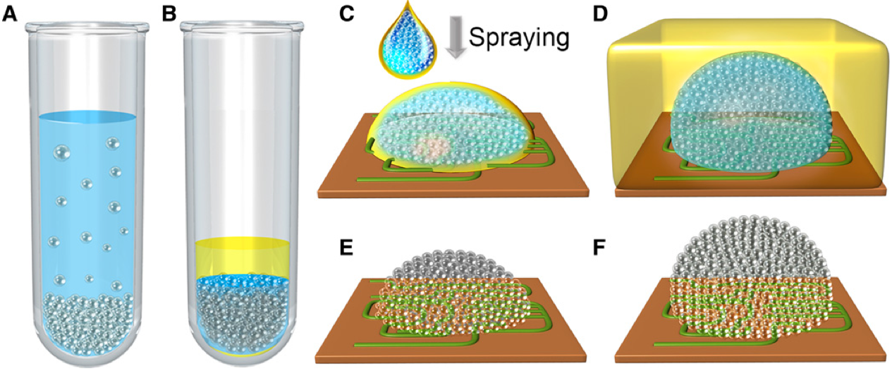
Schematic illustration of the assembly of the all-dielectric TiO2 mSIL. (A) Anatase TiO2 nanoparticles (15 nm) were centrifuged into a tightly packed precipitate. (B) The supernatant was replaced by an organic solvent mixture consisting of hexane and tetrachloroethylene to form a TiO2 nano–solid-fluid. (C) To prepare a hemispherical mSIL, the nano–solid-fluid was directly sprayed onto the sample surface. (D) To prepare a super-hemispherical mSIL, the nano–solid-fluid was sprayed onto the sample surface covered by a thin layer of organic solvent mixture. (E and F) After evaporation of the solvents, the nanoparticles underwent a phase transition to form a more densely packed structure. (credit: Wen Fan et al./Science Advances)

Made of millions of nanobeads, the spheres break up the light beam. Acting as individual minute beams, each bead refracts the light. “We’ve used high-index titanium dioxide (TiO2) nanoparticles as the building element of the lens,” Wang says. “These nanoparticles are able to bend light to a higher degree than water.”

“Each sphere bends the light to a high magnitude and splits the light beam, creating millions of individual beams of light. It is these tiny light beams which enable us to view previously unseen detail.”

Wang believes that the results will be easily replicable and that other labs will soon be adopting the technology and using it for themselves. Titanium dioxide is cheap and readily available, so rather than buying a new microscope, the lenses are applied to the material to be viewed, rather than to the microscope.

“The next challenge is to adapt the technology for use in biology and medicine. This would not require the current use of a combination of dyes and stains and laser light, which change the samples being viewed,” he says.

The lens is described in a paper in the open-access journal Science Advances today (August 12).


Abstract of Three-dimensional all-dielectric metamaterial solid immersion lens for subwavelength imaging at visible frequencies

Although all-dielectric metamaterials offer a low-loss alternative to current metal-based metamaterials to manipulate light at the nanoscale and may have important applications, very few have been reported to date owing to the current nanofabrication technologies. We develop a new “nano–solid-fluid assembly” method using 15-nm TiO2 nanoparticles as building blocks to fabricate the first three-dimensional (3D) all-dielectric metamaterial at visible frequencies. Because of its optical transparency, high refractive index, and deep-subwavelength structures, this 3D all-dielectric metamaterial-based solid immersion lens (mSIL) can produce a sharp image with a super-resolution of at least 45 nm under a white-light optical microscope, significantly exceeding the classical diffraction limit and previous near-field imaging techniques. Theoretical analysis reveals that electric field enhancement can be formed between contacting TiO2 nanoparticles, which causes effective confinement and propagation of visible light at the deep-subwavelength scale. This endows the mSIL with unusual abilities to illuminate object surfaces with large-area nanoscale near-field evanescent spots and to collect and convert the evanescent information into propagating waves. Our all-dielectric metamaterial design strategy demonstrates the potential to develop low-loss nanophotonic devices at visible frequencies.

Anti-inflammatory drug reverses memory loss in Alzheimer’s-disease-model mice

(credit: NIH National Institute on Aging)

Anti-inflammatory drug mefenamic acid completely reversed memory loss and brain inflammation in mice genetically engineered to develop symptoms of Alzheimer’s disease and amyloid beta-induced memory loss, a team led by David Brough, PhD, from the University of Manchester has discovered.

The non-steroidal anti-inflammatory drug (NSAID) drug targets an important inflammatory pathway called the NLRP3 inflammasome, which damages brain cells, according to Brough. This is the first time a drug has been shown to target this inflammatory pathway, highlighting its importance in the disease model, Brough said.

“Because this drug is already available and the toxicity and pharmacokinetics of the drug is known, the time for it to reach patients should, in theory, be shorter than if we were developing completely new drugs. We are now preparing applications to perform early phase II trials to determine a proof-of-concept that the molecules have an effect on neuroinflammation in humans.”

“There is experimental evidence now to strongly suggest that inflammation in the brain makes Alzheimer’s disease worse. Until now, no drug has been available to target this pathway, so we are very excited by this result.”

The research, funded by the Medical Research Council and the Alzheimer’s Society, paves the way for human trials that the team hopes to conduct in the future, but Brough cautions that more research is needed to identify its impact on humans and the long-term implications of its use.

The findings were published Thursday Aug. 11 in an open-access paper authored by Brough and colleagues in the journal Nature Communications.


Abstract of Fenamate NSAIDs inhibit the NLRP3 inflammasome and protect against Alzheimer’s disease in rodent models

Non-steroidal anti-inflammatory drugs (NSAIDs) inhibit cyclooxygenase-1 (COX-1) and COX-2 enzymes. The NLRP3 inflammasome is a multi-protein complex responsible for the processing of the proinflammatory cytokine interleukin-1β and is implicated in many inflammatory diseases. Here we show that several clinically approved and widely used NSAIDs of the fenamate class are effective and selective inhibitors of the NLRP3 inflammasome via inhibition of the volume-regulated anion channel in macrophages, independently of COX enzymes. Flufenamic acid and mefenamic acid are efficacious in NLRP3-dependent rodent models of inflammation in air pouch and peritoneum. We also show therapeutic effects of fenamates using a model of amyloid beta induced memory loss and a transgenic mouse model of Alzheimer’s disease. These data suggest that fenamate NSAIDs could be repurposed as NLRP3 inflammasome inhibitors and Alzheimer’s disease therapeutics.

Harvard, Caltech design mechanical signaling, diodes, logic gates for soft robots

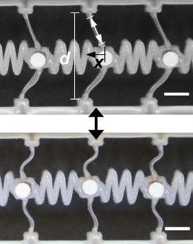
The Harvard/Caltech system for transmitting a mechanical signal consists of a series of bistable elements (the vertical beam, d, shown here) connected by soft coupling elements (wiggly lines), with two stable states. (Top) When a beam is displaced (by amount x), it stores energy. (Bottom) When it snaps back, it releases that stored energy into the coupling element on the right, which continues down the line, like dominos. (Scale bars represent 5 mm.) (credit: Jordan R. Raney/PNAS)

A new way to send mechanical signals through soft robots and other autonomous soft systems has been developed by researchers at the Harvard John A. Paulson School of Engineering and Applied Sciences (SEAS), in collaboration with colleagues at the California Institute of Technology, described in the journal Proceedings of the National Academy of Sciences.

Soft autonomous systems, just like the human body, can perform delicate movements that are safe for humans, unlike mechanical actuators controlled by wires. The problem is that in sending a mechanical signal through a soft material — to make a robot “muscle” move, for example — the signal becomes dissipated (weakened) and dispersed (scattered).

Think tapping on a solid wall to communicate via morse code with someone in the next room vs. tapping out a muffled message on a wall covered with thick, soft foam.

Transmitting signals through soft materials

The researchers solved this problem by using “bistable beams” (structures that function in two distinct states) to store and release elastic energy along the path of a wave.

This new system consists of a chain of bistable elastomeric (rubber-like) beam structures connected by elastomeric linear springs. When a beam is deformed (bent), it snaps and stores energy. As the signal travels along the elastomer, it snaps the beam back into place, releasing the beam’s stored energy and sending the signal downstream, like a line of dominos. This simple bistable system prevents the signal from dissipating downstream.

“This design solves two fundamental problems in transmitting information through materials,” said Katia Bertoldi, the John L. Loeb Associate Professor of the Natural Sciences at SEAS and senior author of the paper.  “It not only overcomes dissipation, but it also eliminates dispersive [spreading out] effects, so that the signal propagates without distortion.  As such, we maintain signal strength and clarity from start to end.” The team used advanced 3D printing techniques to fabricate the system.

Soft diodes and logic gates

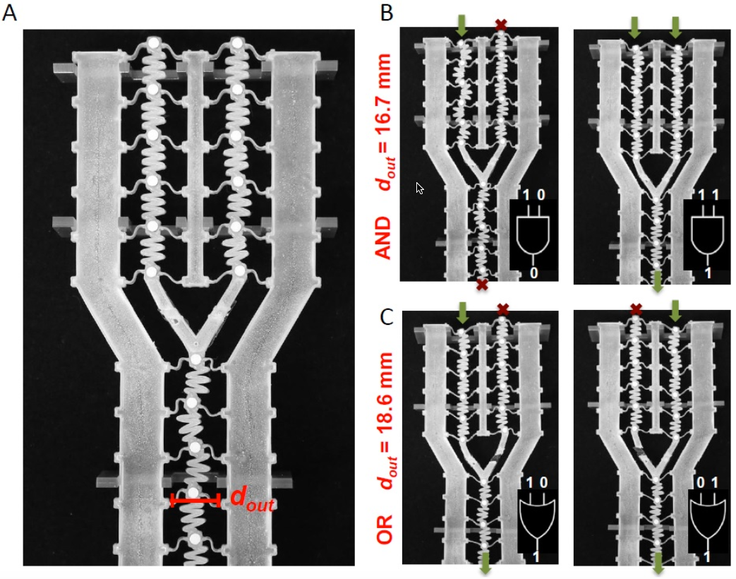
(A) A bifurcated (split into two) signal chain demonstrating tunable logic in a soft mechanical system. The distance d(out) determines the logical behavior, producing either an AND or an OR gate from the same system. (B) When d(out) is small (in this case, 16.7 mm) the energy barrier is higher, so both input signals must be strong to enable the wave to propagate through the output — a logical AND gate; (C) By increasing d(out) (to 18.6 mm in this case), the energy barrier decreases, producing a logical OR gate; in which case, either (or both) input signal has sufficient energy to trigger an output signal. (credit: Jordan R. Raney/PNAS)

The team also took the system a step further, designing and 3D-printing soft diodes and logic gates (a basic computational element that is normally part of a computer chip) using this same signal-transmission design. The gate can be controlled to act either as an AND (both inputs must be present to trigger the gate to fire) or as an OR gate (either one or both will trigger the gate to fire).

This research was supported by the National Science Foundation and the Harvard University Materials Research Science and Engineering Center (MRSEC).


Abstract of Stable propagation of mechanical signals in soft media using stored elastic energy

Soft structures with rationally designed architectures capable of large, nonlinear deformation present opportunities for unprecedented, highly-tunable devices and machines. However, the highly-dissipative nature of soft materials intrinsically limits or prevents certain functions, such as the propagation of mechanical signals. Here, we present an architected soft system comprised of elastomeric bistable beam elements connected by elastomeric linear springs. The dissipative nature of the polymer readily damps linear waves, preventing propagation of any mechanical signal beyond a short distance, as expected. However, the unique architecture of the system enables propagation of stable, nonlinear solitary transition waves with constant, controllable velocity and pulse geometry over arbitrary distances. Since the high damping of the material removes all other linear, small amplitude excitations, the desired pulse propagates with high delity and controllability. This phenomenon can be used to control signals, as demonstrated by the design of soft mechanical diodes and logic gates.

Self-propelling liquid metals: the future of soft electronics?

Liquid metal material with metallic core and semiconducting skin (credit: RMIT University)

Imagine a soft liquid-metal material right out of  the T-1000 Terminator movie character. One that can morph itself into different self-propelling soft electronic circuits that act like live cells, communicating with each other.

Using a liquid metallic core* and semiconducting skin, such a soft material might be used to make instant flexible 3D electronic displays. Or morph into self-propelled biomedical diagnostic sensors, for example, reconfiguring themselves on demand, say RMIT University researchers.

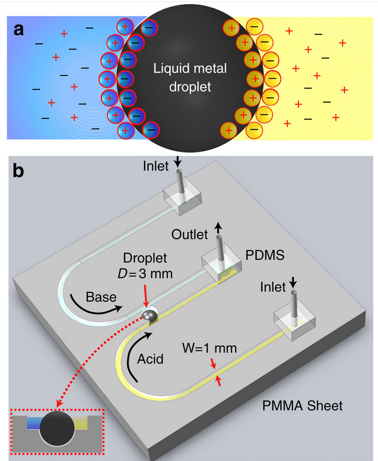
Diagram of ionic-imbalance-induced self-propulsion of liquid metals. (a) Schematic of the droplet and arrangement of ions, forming the EDL. (b) Schematic of the experimental setup showing two U-shaped open-top (see inset) inlet channels, which extend in parallel and join at an outlet. Two channels carry different types of electrolytes (acidic in yellow and basic in blue). Two parallel flows come in contact with the Galinstan droplet (3 mm diameter) residing in a recess. (credit: Ali Zavabeti et al./Nature Communications)

Morphing metal — more like live cells

To achieve that magic, Professor Kourosh Kalantar-zadeh and his group first immersed liquid-metal droplets in water. The droplets were able to move about freely in three dimensions, driven by pH (acid or base) or ionic (electric charge) concentration gradients across a liquid metal droplet, which induced deformation and surface flow.

“We adjusted the concentrations of acid, base, and salt components in the water and investigated the effect. Simply tweaking the water’s chemistry made the liquid metal droplets move and change shape, without any need for external mechanical, electronic, or optical stimulants.”

No, these are not lollipops. These devices demonstrate continuous motion of a self-propelling liquid metal droplet under a pH gradient, shown at different time intervals (left to right). The droplet is placed in a fluidic channel, midway between two reservoirs filled with different acid and base electrolytes. (credit: RMIT University)

The elastic electronic soft circuit systems act more like live cells, moving around autonomously and communicating with each other to form new circuits, rather than being stuck in one configuration.

“Using this discovery, we were able to create moving objects, switches, and pumps that could operate autonomously — self-propelling liquid metals driven by the composition of the surrounding fluid,” Kalantar-zadeh said. “Eventually, using the fundamentals of this discovery, it may be possible to build a 3D liquid metal humanoid on demand.”

The research was published August 4 in open-access Nature Communications.

* Galinstan, an alloy of of 68.5% gallium, 21.5% indium, and 10% tin, is used as the model liquid metal. Galinstan’s melting point can be lowered to below 0 °C (32 °F).


RMIT University | Liquid metals propel future electronics | RMIT University


Abstract of Ionic imbalance induced self-propulsion of liquid metals

Components with self-propelling abilities are important building blocks of small autonomous systems and the characteristics of liquid metals are capable of fulfilling self-propulsion criteria. To date, there has been no exploration regarding the effect of electrolyte ionic content surrounding a liquid metal for symmetry breaking that generates motion. Here we show the controlled actuation of liquid metal droplets using only the ionic properties of the aqueous electrolyte. We demonstrate that pH or ionic concentration gradients across a liquid metal droplet induce both deformation and surface Marangoni flow. We show that the Lippmann dominated deformation results in maximum velocity for the self-propulsion of liquid metal droplets and illustrate several key applications, which take advantage of such electrolyte-induced motion. With this finding, it is possible to conceive the propulsion of small entities that are constructed and controlled entirely with fluids, progressing towards more advanced soft systems.

Ultrasonic wireless ‘neural dust’ sensors monitor nerves, muscles in real time

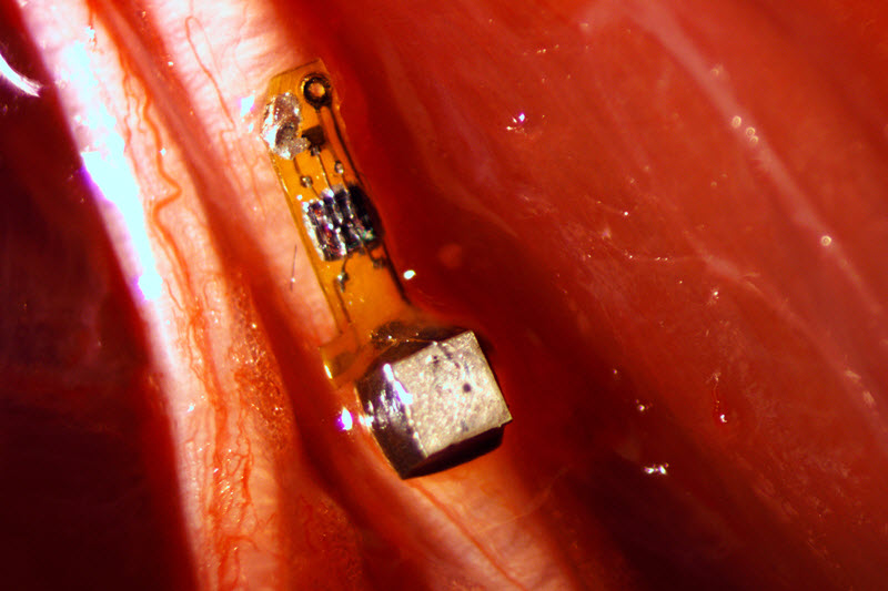
Prototype wireless battery-less “neural dust” mote (3 x 1 x 1 millimeters) with electrodes attached to a nerve fiber in a rat. The mote contains a piezoelectric crystal (silver cube) that converts ultrasonic signals to electrical current, powering a simple electronic circuit containing a transistor (black square) that responds to the voltage generated by a nerve firing and triggers the piezoelectric crystal to create ultrasonic backscatter, which indicates detection of a neural signal. (photo credit: Ryan Neely/UC Berkeley)

University of California, Berkeley engineers have designed and built millimeter-scale device wireless, batteryless “neural dust” sensors and implanted them in muscles and peripheral nerves of rats to make in vivo electrophysiological recordings.

The new technology opens the door to “electroceuticals” — bioelectronic methods to monitor and record wireless electromyogram (EMG) signals from muscle membranes and electroneurogram (ENG) signals from local neuron electrical activity, and to stimulate the immune system, reduce inflammation, and treat disorders such as epilepsy.

The technology could also improve neural control of prosthetics (allowing a paraplegic to control a computer or a robotic arm, for example) by stimulating nerves and muscles directly, instead of requiring implanted wires.

The neural-dust sensors use ultrasound technology to both power the sensors and read out measurements. Ultrasound is already well-developed for hospital use and can penetrate nearly anywhere in the body, unlike radio waves.

The researchers reported their findings August 3 in an open-access paper in the journal Neuron.

How a neural dust “mote” sensor monitors neural and muscle signals

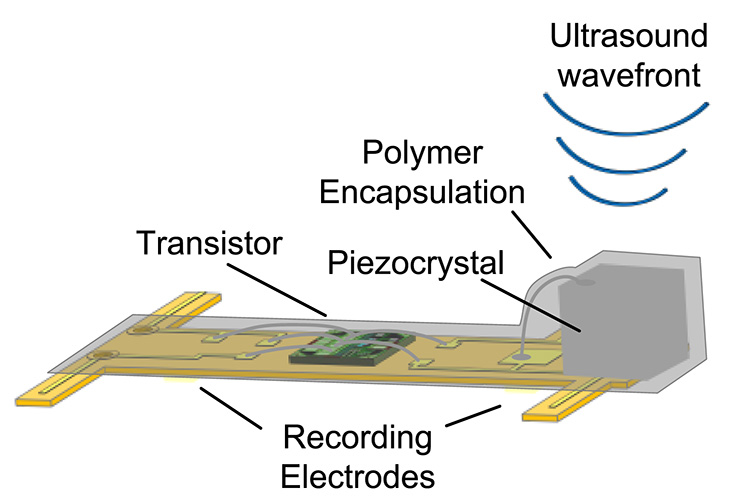
Diagram showing the components of the neural-dust mote (sensor). The entire device is covered in a biocompatible gel. (credit: Dongjin Seo et al./Neuron)

1. A team implants the neural dust mote. In the reported study, the mote was implanted in the rat sciatic nerve to do ENG recordings and in the gastrocnemius muscle to do EMG recordings. The tether-less connection also avoids potential infections and adverse biological responses due to micro-motion of the implant within the tissue.

2. An external ultrasonic generator sends a ultrasound signal to a piezoelectric crystal, which converts the sound energy into an electrical voltage, used to power a transistor circuit — no battery required.

3. When neurons or muscle fibers fire, they generate a tiny voltage (action potential) that the two electrodes pick up and send to the transistor.

4. The transistor amplifies the signal and drives the piezoelectric crystal to vibrate at an ultrasonic rate.

5. That vibration interferes with the transmitted ultrasonic signal, causing a modified “backscatter” signal that communicates information about the voltage across the sensor’s two electrodes.

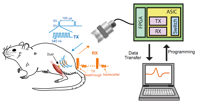
In vivo experimental setup for a tether-less neural dust electromyography (EMG) recording from the gastrocnemius muscle in rats. The neural dust mote was placed on the exposed muscle surface, and the wound was closed with surgical suture. The ASIC chip switches between ultrasound transmit and receive modules.Triggered by the FPGA chip, the external transducer sends ultrasound (TX, green) to the mote. The backscatter received signal (RX, red) carries information about the EMG voltage that is decoded and recorded/displayed on a computer. The dust mote was pinged every 100 microseconds with six 540-nanosecond ultrasound pulses. (credit: Dongjin Seo et al./Neuron)

6. The backscatter ultrasound signal is decoded to extract EMG or ENG data.

7. A computer displays and records the information.

Microscale motes: future research

The experiments so far have involved the peripheral nervous system and muscles, using an external ultrasonic patch over the implanted site to acquire  information from the motes for the desired diagnosis or therapy.

But according to the researchers, neural dust motes can be implanted anywhere in the body, including the central nervous system and brain to control prosthetics. This would be an alternative to today’s implantable electrodes (for Parkinson’s disease, for example), which require wires that pass through holes in the skull and degrade within one to two years.

The researchers are now building motes from biocompatible thin films, which would potentially last in the body without degradation for a decade or more. Up to hundreds of wireless sensors could be sealed in, avoiding infection and unwanted movement of the electrodes, and could last a timeline, according to the researchers.

The team is also now working to miniaturize the device further and they plan to use beam-steering technology to focus the ultrasonic signals on individual motes. The team is also building little backpacks for rats to hold the ultrasound transceiver that will record data from implanted motes. And the researchers are working to expand the motes’ ability to detect non-electrical signals, such as oxygen or hormone levels.

The researchers estimate that they could eventually shrink the sensors down to a cube 50 micrometers on a side. At that size, the motes could monitor a few specific nerve axons and continually record their electrical activity.

The researchers conceived of the idea of neural dust about five years ago, but initial attempts to power an implantable device and read out the data using radio waves were disappointing. Radio attenuates very quickly with distance in tissue, so communicating with devices deep in the body would be difficult without using potentially damaging high-intensity radiation. In 2013, the researchers published an open-access arXiv paper that described how a neural-dust system with ultrasonic signals might work.

The ongoing research is supported by the U.S. Defense Advanced Research Projects Agency as part of DARPA’s Electrical Prescriptions (ElectRx) program, which is focused in part on developing interface technologies that are suitable for chronic use for biosensing and neuromodulation of specific peripheral nerves.


UC Berkeley | “Neural dust” sensor


Abstract of Wireless Recording in the Peripheral Nervous System with Ultrasonic Neural Dust

The emerging field of bioelectronic medicine seeks methods for deciphering and modulating electrophysiological activity in the body to attain therapeutic effects at target organs. Current approaches to interfacing with peripheral nerves and muscles rely heavily on wires, creating problems for chronic use, while emerging wireless approaches lack the size scalability necessary to interrogate small-diameter nerves. Furthermore, conventional electrode-based technologies lack the capability to record from nerves with high spatial resolution or to record independently from many discrete sites within a nerve bundle. Here, we demonstrate neural dust, a wireless and scalable ultrasonic backscatter system for powering and communicating with implanted bioelectronics. We show that ultrasound is effective at delivering power to mm-scale devices in tissue; likewise, passive, battery-less communication using backscatter enables high-fidelity transmission of electromyogram (EMG) and electroneurogram (ENG) signals from anesthetized rats. These results highlight the potential for an ultrasound-based neural interface system for advancing future bioelectronics-based therapies.

No, exercise does not wipe out previous memories

2016 Rio Games Opening Ceremony promo (credit: NBC)

A week ago on KurzweilAI, we learned that prolonged sitting may increase risk of death, but that an hour of moderate exercise a day is enough to counter health risks. Now new research suggests that such exercise results in larger brain size and lowered dementia risk, while other new research suggests that the new neurons created in that exercise preserve old memories, contrary to previous research.

Exercise results in larger brain size and lowered dementia risk

UCLA researchers have found an association between low physical activity and a higher risk for dementia in older individuals, based on data from the landmark Framingham Heart Study.

The researchers found that physical activity particularly affected the size of the hippocampus, involved in short-term memory. They also found the protective effect of regular physical activity against dementia was strongest in people age 75 and older.

This suggests that regular physical activity for older adults could lead to higher brain volumes and a reduced risk for developing dementia.

The Framingham study was begun in 1948 primarily as a way to trace factors and characteristics leading to cardiovascular disease, but also examining dementia and other physiological conditions. For this study, the UCLA researchers followed an older, community-based cohort from the Framingham study for more than a decade to examine the association between physical activity and the risk for incident dementia and subclinical brain MRI markers of dementia.

The study appears in the Journals of Gerontology Series A: Biological Sciences and Medical Sciences. It was supported by an NIH/National Heart, Lung, and Blood Institute contract and training grant, the National Institute on Aging, the National Institute of Neurological Disorders and Stroke, and the American Heart Association.

New neurons created through exercise don’t cause you to forget old memories

Meanwhile, Texas A&M College of Medicine scientists have found in a study recently published in the Journal of Neuroscience that exercise causes more new neurons to be formed in a critical brain region — and contrary to an earlier study, these new neurons do not cause the individual to forget old memories.

Exercise is well known for its cognitive benefits, thought to occur because it causes neurogenesis, or the creation of new neurons, in the hippocampus, which is a key brain region for learning, memory and mood regulation. So it was a surprise in 2014 when a research study, published in the journal Science, found that exercise caused mice to forget what they’d already learned.

“It stunned the field of hippocampal neurogenesis,” said Ashok K. Shetty, PhD, a professor in the Texas A&M College of Medicine Department of Molecular and Cellular Medicine, associate director of the Institute for Regenerative Medicine, and research career scientist at the Central Texas Veterans Health Care System.

The animal models in the exercise group — in the previous study — showed far more neurogenesis than the control group, but these additional neurons seemed to erase memories that were formed before they started the exercise regimen. To test this, the researchers removed the extra neurons, and the mice suddenly were able to remember again.

Replicating the research with rats reversed the outcome

Shetty and his team decided to replicate this earlier research, using rats instead of mice. Rats are thought to be more like humans physiologically, with more-similar neuronal workings. They found that these animal models showed no such degradation in memories.

The researchers trained their animal models to complete a task over the course of four days, followed by several days of memory consolidation by performing the task over and over again. Then, half the trained animal models were put into cages with running wheels for several weeks, while the control group remained sedentary.

The rats who ran further over the course of that time had much greater neurogenesis in their hippocampus, and all rats who had access to a wheel (and therefore ran at least some), had greater neurogenesis than the sedentary group.

Importantly, despite differing levels of increased neurogenesis, both moderate runners and brisk runners (those who ran further than average) in Shetty’s study showed the same ability as the sedentary runners to recall the task they learned before they began to exercise.

This means even a large amount of running (akin to people who perform significant amount of exercise on a daily basis) doesn’t interfere with the recall of memory.

Why fidgeting can protect leg arteries and improve learning

(credit: Dave Clark/National Education Association)

But not everybody has that extra hour — or is motivated to exercise. For all of us couch/desk-bound folks, new research suggests a simple sitting exercise that may tide you over until you can go for a walk or run: fidgeting.

Ignore that teacher advice to “sit in your seat and don’t fidget!” — which the National Education Association says in this article actually improves learning — and suggests footrests using old tires to make giant rubber bands to facilitate fidgeting.

University of Missouri researchers have found that fidgeting while sitting can also protect the arteries in legs and potentially help prevent the arterial disease that may be brought on by binge TV watching or working at a computer.

In the study, the researchers compared the leg vascular function of 11 healthy young men and women before and after three hours of sitting. While sitting, the participants were asked to fidget one leg intermittently, tapping one foot for one minute and then resting it for four minutes, while the other leg remained still throughout. On average, the participants moved their feet 250 times per minute.

The researchers then measured the blood flow of the popliteal — an artery in the lower leg — and found that the fidgeting leg had a significant increase in blood flow, as expected, while the stationary leg experienced a reduction in blood flow.

Research has shown that increased blood flow and its associated shear stress — the friction of the flowing blood on the artery wall — is an important stimulus for vascular health.

While only one leg was exposed to fidgeting during the experiment, in a real-world scenario the researchers recommend tapping both legs to maximize the beneficial effects.

But not a substitute for walking and exercise

“Many of us sit for hours at a time, whether it’s binge watching our favorite TV show or working at a computer,” said Jaume Padilla, PhD, an assistant professor of nutrition and exercise physiology at MU and lead author of the study.

“We wanted to know whether a small amount of leg fidgeting could prevent a decline in leg vascular function caused by prolonged sitting. While we expected fidgeting to increase blood flow to the lower limbs, we were quite surprised to find this would be sufficient to prevent a decline in arterial function.”

But fidgeting is not a substitute for walking and exercise, which produce more overall cardiovascular benefits, the researchers caution.

“You should attempt to break up sitting time as much as possible by standing or walking,” Padilla said. “But if you’re stuck in a situation in which walking just isn’t an option, fidgeting can be a good alternative. Any movement is better than no movement.”

The study was recently published in the American Journal of Physiology Heart and Circulatory Physiology and was supported by the NIH and the Japan Society for the Promotion of Science.


Abstract of Physical Activity, Brain Volume, and Dementia Risk: The Framingham Study

Background: Several longitudinal studies found an inverse relationship between levels of physical activity and cognitive decline, dementia, and/or Alzheimer’s disease (AD), but results have been inconsistent. We followed an older, community-based cohort for over a decade to examine the association of physical activity with the risk of incident dementia and subclinical brain MRI markers of dementia.

Methods: The physical activity index (PAI) was assessed in the Framingham Study Original and Offspring cohorts, aged 60 years or older. We examined the association between PAI and risk of incident all-cause dementia and AD in participants of both cohorts who were cognitively intact and had available PAI (n = 3,714; 54% women; mean age = 70±7 years). We additionally examined the association between PAI and brain MRI in the Offspring cohort (n = 1,987).

Results: Over a decade of follow-up, 236 participants developed dementia (188 AD). Participants in the lowest quintile of PAI had an increased risk of incident dementia compared with those in higher quintiles (hazard ratio [HR] = 1.50, 95% confidence interval [CI] = 1.04–1.97, p = .028) in a multivariable-adjusted model. Secondary analysis revealed that this relation was limited to participants who were apolipoprotein (APO)E ε4 allele noncarriers (HR = 1.58, 95% CI = 1.08–2.32; p = .018) and strongest in participants aged 75 years or older. PAI was also linearly related to total brain and hippocampal volumes (β ± SE = 0.24±0.06; p < .01 and 0.004±0.001; p = .003, respectively).

Conclusion: Low physical activity is associated with a higher risk for dementia in older individuals, suggesting that a reduced risk of dementia and higher brain volumes may be additional health benefits of maintaining physical activity into old age.


Abstract of Voluntary Running Exercise-Mediated Enhanced Neurogenesis Does Not Obliterate Retrograde Spatial Memory

Running exercise (RE) improves cognition, formation of anterograde memories, and mood, alongside enhancing hippocampal neurogenesis. A previous investigation in a mouse model showed that RE-induced increased neurogenesis erases retrograde memory (Akers et al., 2014). However, it is unknown whether RE-induced forgetting is common to all species. We ascertained whether voluntary RE-induced enhanced neurogenesis interferes with the recall of spatial memory in rats. Young rats assigned to either sedentary (SED) or running exercise (RE) groups were first subjected to eight learning sessions in a water maze. A probe test (PT) conducted 24 h after the final training session confirmed that animals in either group had a similar ability for the recall of short-term memory. Following this, rats in the RE group were housed in larger cages fitted with running wheels, whereas rats in the SED group remained in standard cages. Animals in the RE group ran an average of 78 km in 4 weeks. A second PT performed 4 weeks after the first PT revealed comparable ability for memory recall between animals in the RE and SED groups, which was evidenced through multiple measures of memory retrieval function. The RE group displayed a 1.5- to 2.1-fold higher hippocampal neurogenesis than SED rats. Additionally, both moderate and brisk RE did not interfere with the recall of memory, although increasing amounts of RE proportionally enhanced neurogenesis. In conclusion, RE does not impair memory recall ability in a rat model despite substantially increasing neurogenesis.

SIGNIFICANCE STATEMENT Running exercise (RE) improves new memory formation along with an increased neurogenesis in the hippocampus. In view of a recent study showing that RE-mediated increased hippocampal neurogenesis promotes forgetfulness in a mouse model, we ascertained whether a similar adverse phenomenon exists in a rat model. Memory recall ability examined 4 weeks after learning confirmed that animals that had run a mean of 78 km and displayed a 1.5- to 2.1-fold increase in hippocampal neurogenesis demonstrated similar proficiency for memory recall as animals that had remained sedentary. Furthermore, both moderate and brisk RE did not interfere with memory recall, although increasing amounts of RE proportionally enhanced neurogenesis, implying that RE has no adverse effects on memory recall.


Abstract of Prolonged sitting-induced leg endothelial dysfunction is prevented by fidgeting

Prolonged sitting impairs endothelial function in the leg vasculature, and this impairment is thought to be largely mediated by a sustained reduction in blood flow-induced shear stress. Indeed, preventing the marked reduction of shear stress during sitting with local heating abolishes the impairment in popliteal artery endothelial function. Herein, we tested the hypothesis that sitting-induced reductions in shear stress and ensuing endothelial dysfunction would be prevented by periodic leg movement, or “fidgeting.” In 11 young, healthy subjects, bilateral measurements of popliteal artery flow-mediated dilation (FMD) were performed before and after a 3-h sitting period during which one leg was subjected to intermittent fidgeting (1 min on/4 min off) while the contralateral leg remained still throughout and served as an internal control. Fidgeting produced a pronounced increase in popliteal artery blood flow and shear rate (prefidgeting, 33.7 ± 2.6 s−1 to immediately postfidgeting, 222.7 ± 28.3 s−1; mean ± SE; P < 0.001) that tapered off during the following 60 s. Fidgeting did not alter popliteal artery blood flow and shear rate of the contralateral leg, which was subjected to a reduction in blood flow and shear rate throughout the sitting period (presit, 71.7 ± 8.0 s−1 to 3-h sit, 20.2 ± 2.9 s−1; P < 0.001). Popliteal artery FMD was impaired after 3 h of sitting in the control leg (presit, 4.5 ± 0.3% to postsit: 1.6 ± 1.1%; P = 0.039) but improved in the fidgeting leg (presit, 3.7 ± 0.6% to postsit, 6.6 ± 1.2%; P = 0.014). Collectively, the present study provides evidence that prolonged sitting-induced leg endothelial dysfunction is preventable with small amounts of leg movement while sitting, likely through the intermittent increases in vascular shear stress.

IBM scientists emulate neurons with phase-change technology

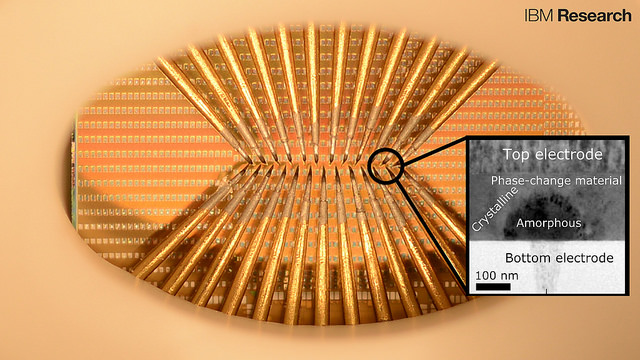
A prototype chip with large arrays of phase-change devices that store the state of artificial neuronal populations in their atomic configuration. The devices are accessed via an array of probes in this prototype to allow for characterization and testing. The tiny squares are contact pads used to access the nanometer-scale phase-change cells (inset).  Each set of probes can access a population of 100 cells. There are thousands to millions of these cells on one chip and IBM accesses them (in this particular photograph) by means of the sharp needles (probe card). (credit: IBM Research)

Scientists at IBM Research in Zurich have developed artificial neurons that emulate how neurons spike (fire). The goal is to create energy-efficient, high-speed, ultra-dense integrated neuromorphic (brain-like) technologies for applications in cognitive computing, such as unsupervised learning for detecting and analyzing patterns.

Applications could include internet of things sensors that collect and analyze volumes of weather data for faster forecasts and detecting patterns in financial transactions, for example.

The results of this research appeared today (Aug. 3) as a cover story in the journal Nature Nanotechnology.

Emulating neuron spiking

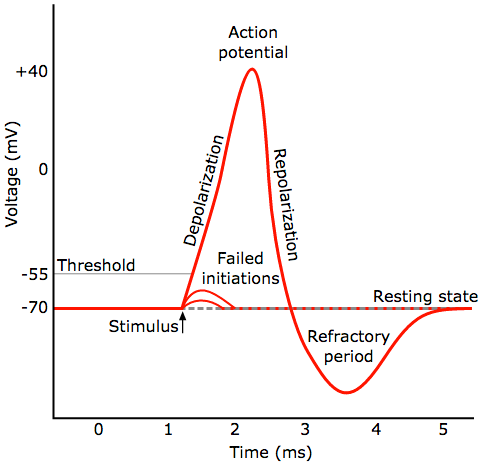
General pattern of a neural spike (action potential). A neuron fires (generates a rapid action potential, or voltage, when triggered by a stimulus) a signal from a synapse (credit: Chris 73/Diberri CC)

IBM’s new neuron-like spiking mechanism is based on a recent IBM breakthrough in phase-change materials. Phase-change materials are used for storing and processing digital data in re-writable Blu-ray discs, for example. The new phase-change materials developed by IBM recently are used instead for storing and processing analog data — like the synapses and neurons in our biological brains.

The new phase-change materials also overcome  problems in conventional computing, where there’s a separate memory and logic unit, slowing down computation. These functions are combined in the new artificial neurons, just as they are in a biological neuron.

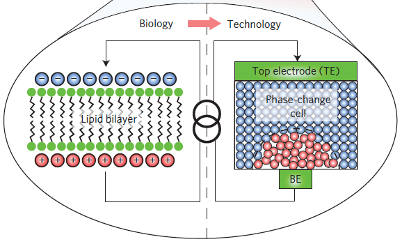
In biological neurons, a thin lipid-bilayer membrane separates the electrical charges inside the cell from those outside it. The membrane potential is altered by the arrival of excitatory and inhibitory postsynaptic potentials through the dendrites of the neuron, and upon sufficient excitation of the neuron (a phase change), an action potential, or spike, is generated. IBM’s new germanium-antimony-tellurium (GeSbTe or GST) phase-change material emulates this process. It has two stable states: an amorphous one (without a clearly defined structure) and a crystalline one (with a structure). (credit: Tomas Tuma et al./Nature Nanotechnology)

Alternative to von-Neumann-based algorithms

In addition, previous attempts to build artificial neurons are built using CMOS-based circuits, the standard transistor technology we have in our computers. The new phase-change technology can reproduce similar functionality at reduced power consumption. The artificial neurons are also superior in functioning at nanometer-length-scale dimensions and feature native stochasticity (based on random variables, simulating neurons).

“Populations of stochastic phase-change neurons, combined with other nanoscale computational elements such as artificial synapses, could be a key enabler for the creation of a new generation of extremely dense neuromorphic computing systems,” said Tomas Tuma, a co-author of the paper.

“The relatively complex computational tasks, such as Bayesian inference, that stochastic neuronal populations can perform with collocated processing and storage render them attractive as a possible alternative to von-Neumann-based algorithms in future cognitive computers,” the IBM scientists state in the paper.

IBM scientists have organized hundreds of these artificial neurons into populations and used them to represent fast and complex signals. These artificial neurons have been shown to sustain billions of switching cycles, which would correspond to multiple years of operation at an update frequency of 100 Hz. The energy required for each neuron update was less than five picojoule and the average power less than 120 microwatts — for comparison, 60 million microwatts power a 60 watt lightbulb.


IBM Research | All-memristive neuromorphic computing with level-tuned neurons


Abstract of Stochastic phase-change neurons

Artificial neuromorphic systems based on populations of spiking neurons are an indispensable tool in understanding the human brain and in constructing neuromimetic computational systems. To reach areal and power efficiencies comparable to those seen in biological systems, electroionics-based and phase-change-based memristive devices have been explored as nanoscale counterparts of synapses. However, progress on scalable realizations of neurons has so far been limited. Here, we show that chalcogenide-based phase-change materials can be used to create an artificial neuron in which the membrane potential is represented by the phase configuration of the nanoscale phase-change device. By exploiting the physics of reversible amorphous-to-crystal phase transitions, we show that the temporal integration of postsynaptic potentials can be achieved on a nanosecond timescale. Moreover, we show that this is inherently stochastic because of the melt-quench-induced reconfiguration of the atomic structure occurring when the neuron is reset. We demonstrate the use of these phase-change neurons, and their populations, in the detection of temporal correlations in parallel data streams and in sub-Nyquist representation of high-bandwidth signals.

Eating more plant protein associated with lower risk of death

Some high-protein plant sources (credit: iStock)

Eating more protein from plant sources was associated with a lower risk of death, while eating more protein from animals was associated with a higher risk of death — especially among adults with at least one unhealthy behavior such as smoking, drinking, and being overweight or sedentary — according to an open-access survey article published online by JAMA Internal Medicine.

Mingyang Song, M.D., Sc.D., of Massachusetts General Hospital and Harvard Medical School, and coauthors used data from two large U.S. studies, with repeated measures of diet through food questionnaires and up to 32 years of follow-up of 131,342 participants.

The authors report:

  • After adjusting for major lifestyle and dietary risk factors, every 10 percent increment of animal protein from total calories was associated with a 2 percent higher risk of death from all causes and an 8 percent increased risk of death from cardiovascular disease death.
  • In contrast, eating more plant protein was associated with a 10 percent lower risk of death from all causes for every 3 percent increment of total calories and a 12 percent lower risk of cardiovascular death.
  • Increased mortality associated with eating more animal protein was more pronounced among study participants who were obese and those who drank alcohol heavily.
  • The association between eating more plant protein and lower mortality was stronger among study participants who smoked, drank at least 14 grams of alcohol a day, were overweight or obese, were physically inactive or were younger than 65 or older than 80.
  • Substituting 3 percent of calories from animal protein with plant protein was associated with a lower risk of death from all causes: 34 percent for replacing processed red meat, 12 percent for replacing unprocessed red meat and 19 percent for replacing eggs.

“Substitution of plant protein for animal protein, especially from processed red meat, may confer substantial health benefit. Therefore, public health recommendations should focus on improvement of protein sources,” the study concludes.

Limitations of the study

Limitations include the study’s observational design, so residual confounding (other mitigating factors) cannot be excluded, and “the moderately higher protein consumption (median, 19% of calories) in our study population compared with the general U.S. population (15%-16%), thus limiting our ability to assess the effect of the very low end of intake,” the authors note. “Other components in protein-rich foods (e.g., sodium,nitrates, and nitrites in processed red meat), in addition to protein per se, may have a critical health effect.”

Some nutritionists also advise that deficiencies in the diet of vegetarians may include vitamin B(12), vitamin D, omega-3 fatty acids, calcium, iron, and zinc, which may require supplements and fortified foods, according to a 2010 analysis in the journal Nutrition in Clinical Practice.

* They examined hazard ratios (risk) for all-cause and cause-specific mortality in relation to eating animal protein vs. plant protein. Median protein intake, measured as a percentage of calories, was 14 percent for animal protein and 4 percent for plant protein. Among 131,342 study participants, 85,013 (64.7 percent) were women and the average age of participants was 49.


Abstract of Association of Animal and Plant Protein Intake With All-Cause and Cause-Specific Mortality

Importance  Defining what represents a macronutritionally balanced diet remains an open question and a high priority in nutrition research. Although the amount of protein may have specific effects, from a broader dietary perspective, the choice of protein sources will inevitably influence other components of diet and may be a critical determinant for the health outcome.

Objective  To examine the associations of animal and plant protein intake with the risk for mortality.

Design, Setting, and Participants  This prospective cohort study of US health care professionals included 131 342 participants from the Nurses’ Health Study (1980 to end of follow-up on June 1, 2012) and Health Professionals Follow-up Study (1986 to end of follow-up on January 31, 2012). Animal and plant protein intake was assessed by regularly updated validated food frequency questionnaires. Data were analyzed from June 20, 2014, to January 18, 2016.

Main Outcomes and Measures  Hazard ratios (HRs) for all-cause and cause-specific mortality.

Results  Of the 131 342 participants, 85 013 were women (64.7%) and 46 329 were men (35.3%) (mean [SD] age, 49 [9] years). The median protein intake, as assessed by percentage of energy, was 14% for animal protein (5th-95th percentile, 9%-22%) and 4% for plant protein (5th-95th percentile, 2%-6%). After adjusting for major lifestyle and dietary risk factors, animal protein intake was weakly associated with higher mortality, particularly cardiovascular mortality (HR, 1.08 per 10% energy increment; 95% CI, 1.01-1.16; P for trend = .04), whereas plant protein was associated with lower mortality (HR, 0.90 per 3% energy increment; 95% CI, 0.86-0.95; P for trend < .001). These associations were confined to participants with at least 1 unhealthy lifestyle factor based on smoking, heavy alcohol intake, overweight or obesity, and physical inactivity, but not evident among those without any of these risk factors. Replacing animal protein of various origins with plant protein was associated with lower mortality. In particular, the HRs for all-cause mortality were 0.66 (95% CI, 0.59-0.75) when 3% of energy from plant protein was substituted for an equivalent amount of protein from processed red meat, 0.88 (95% CI, 0.84-0.92) from unprocessed red meat, and 0.81 (95% CI, 0.75-0.88) from egg.

Conclusions and Relevance  High animal protein intake was positively associated with mortality and high plant protein intake was inversely associated with mortality, especially among individuals with at least 1 lifestyle risk factor. Substitution of plant protein for animal protein, especially that from processed red meat, was associated with lower mortality, suggesting the importance of protein source.

Miniature portable device produces biopharmaceuticals on demand at point-of-care

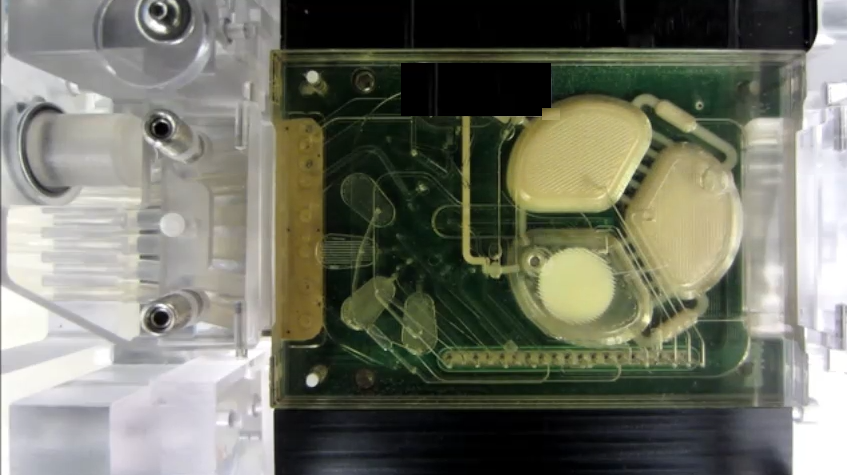
The new portable production system is designed to manufacture a range of biopharmaceuticals on-demand. The microbioreactor contains a polycarbonate-PDMS membrane-polycarbonate sandwiched chip with active microfluidic components (thin white lines) that are equipped for pneumatic routing of reagents, precise peristaltic injection, growth chamber mixing, and fluid extraction. (credit: Pablo Perez-Pinera et al./Nature Communications)

MIT researchers with DARPA funding have developed a portable device for manufacturing a range of biopharmaceuticals on demand, virtually anywhere.

For medics on the battlefield and doctors in remote or developing parts of the world, getting rapid access to the drugs needed to treat patients can be challenging. That’s because biopharmaceutical drugs, which are used in a wide range of therapies including vaccines and treatments for diabetes and cancer, are currently produced in large, centralized fermentation plants. Then they must be transported to the treatment site, which can be expensive, time-consuming, and difficult to execute in areas with poor supply chains.

In an open-access paper published Friday July 29 in the journal Nature Communications, the researchers demonstrate that the system can be used to produce a single dose of treatment from a compact device containing just a small droplet of cells in a liquid.

The system could ultimately be carried onto the battlefield and used to produce treatments at the point of care. It could also be used to manufacture a vaccine to prevent a disease outbreak in a remote village, according to senior author Tim Lu, an associate professor of biological engineering and electrical engineering and computer science, and head of the Synthetic Biology Group at MIT’s Research Laboratory of Electronics. “Imagine you were on Mars or in a remote desert, without access to a full formulary; you could program the yeast to produce drugs on demand locally,” Lu says.

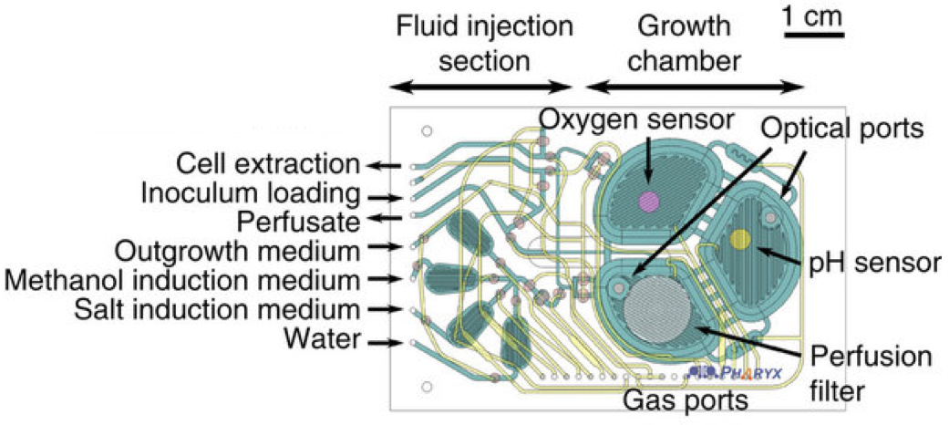
The microbioreactor contains microfluidic circuits (green), sensors for monitoring oxygen and acidity, and a filter to retain the cells while the therapeutic protein is extracted. (credit: Pablo Perez-Pinera et al./Nature Communications)

The prototype system is based on a programmable strain of yeast, Pichia pastoris, which can be induced to express (generate) one of two therapeutic proteins when exposed to a particular chemical trigger. The researchers chose P. pastoris because it can grow to very high densities on simple and inexpensive carbon sources, and is able to express large amounts of protein. “We altered the yeast so it could be more easily genetically modified, and could include more than one therapeutic in its repertoire,” Lu says.

In an experiment, when the researchers exposed the modified yeast to estrogen β-estradiol, the cells expressed recombinant human growth hormone (rHGH). But when they exposed the same cells to methanol, the yeast expressed the protein interferon.

How to create a DIY biopharmaceutical drug

(credit: Pablo Perez-Pinera et al./Nature Communications)

  1. Put the yeast cells into the millimeter-scale table-top microbioreactor.
  2. Feed a  liquid containing the desired chemical trigger (such as methanol) into the reactor to mix with the cells.
  3. Gently massage the liquid droplet to ensure its contents are fully mixed together.
  4. Pressurize the gas in the reactor. That causes oxygen to flow through a silicone rubber membrane and allows carbon dioxide to be extracted.
  5. The device continuously monitors conditions within microfluidic chip, including monitors cell density, oxygen levels, temperature, and pH, to ensure the optimum environment for cell growth.
  6. If you need a different protein, just flush the liquid through a filter, leaving the cells behind*. Then add fresh liquid containing a new chemical trigger to stimulate production of the next protein.

The researchers are now investigating how to use the system in combinatorial treatments, in which multiple therapeutics, such as antibodies, are used together. Combining multiple therapeutics in this way can be expensive if each requires its own production line, Lu says. “But if you could engineer a single strain, or maybe even a consortia of strains that grow together, to manufacture combinations of biologics or antibodies, that could be a very powerful way of producing these drugs at a reasonable cost,” he says.

The portable microbioreactor system operating in an ambulance (credit: Pablo Perez-Pinera et al./Nature Communications)

* Other research teams have previously attempted to build microbioreactors, but these could not retain the protein-producing cells while flushing out the liquid they are mixed with.


Abstract of Synthetic biology and microbioreactor platforms for programmable production of biologics at the point-of-care

Current biopharmaceutical manufacturing systems are not compatible with portable or distributed production of biologics, as they typically require the development of single biologic-producing cell lines followed by their cultivation at very large scales. Therefore, it remains challenging to treat patients in short time frames, especially in remote locations with limited infrastructure. To overcome these barriers, we developed a platform using genetically engineered Pichia pastoris strains designed to secrete multiple proteins on programmable cues in an integrated, benchtop, millilitre-scale microfluidic device. We use this platform for rapid and switchable production of two biologics from a single yeast strain as specified by the operator. Our results demonstrate selectable and near-single-dose production of these biologics in <24 h with limited infrastructure requirements. We envision that combining this system with analytical, purification and polishing technologies could lead to a small-scale, portable and fully integrated personal biomanufacturing platform that could advance disease treatment at point-of-care.

Miniature portable device produces biopharmaceuticals on demand at point-of-care

The new portable production system is designed to manufacture a range of biopharmaceuticals on-demand. The microbioreactor contains a polycarbonate-PDMS membrane-polycarbonate sandwiched chip with active microfluidic components (thin white lines) that are equipped for pneumatic routing of reagents, precise peristaltic injection, growth chamber mixing, and fluid extraction. (credit: Pablo Perez-Pinera et al./Nature Communications)

MIT researchers with DARPA funding have developed a portable device for manufacturing a range of biopharmaceuticals on demand, virtually anywhere.

For medics on the battlefield and doctors in remote or developing parts of the world, getting rapid access to the drugs needed to treat patients can be challenging. That’s because biopharmaceutical drugs, which are used in a wide range of therapies including vaccines and treatments for diabetes and cancer, are currently produced in large, centralized fermentation plants. Then they must be transported to the treatment site, which can be expensive, time-consuming, and difficult to execute in areas with poor supply chains.

In an open-access paper published Friday July 29 in the journal Nature Communications, the researchers demonstrate that the system can be used to produce a single dose of treatment from a compact device containing just a small droplet of cells in a liquid.

The system could ultimately be carried onto the battlefield and used to produce treatments at the point of care. It could also be used to manufacture a vaccine to prevent a disease outbreak in a remote village, according to senior author Tim Lu, an associate professor of biological engineering and electrical engineering and computer science, and head of the Synthetic Biology Group at MIT’s Research Laboratory of Electronics. “Imagine you were on Mars or in a remote desert, without access to a full formulary; you could program the yeast to produce drugs on demand locally,” Lu says.

The microbioreactor contains microfluidic circuits (green), sensors for monitoring oxygen and acidity, and a filter to retain the cells while the therapeutic protein is extracted. (credit: Pablo Perez-Pinera et al./Nature Communications)

The prototype system is based on a programmable strain of yeast, Pichia pastoris, which can be induced to express (generate) one of two therapeutic proteins when exposed to a particular chemical trigger. The researchers chose P. pastoris because it can grow to very high densities on simple and inexpensive carbon sources, and is able to express large amounts of protein. “We altered the yeast so it could be more easily genetically modified, and could include more than one therapeutic in its repertoire,” Lu says.

In an experiment, when the researchers exposed the modified yeast to estrogen β-estradiol, the cells expressed recombinant human growth hormone (rHGH). But when they exposed the same cells to methanol, the yeast expressed the protein interferon.

How to create a DIY biopharmaceutical drug

(credit: Pablo Perez-Pinera et al./Nature Communications)

  1. Put the yeast cells into the millimeter-scale table-top microbioreactor.
  2. Feed a  liquid containing the desired chemical trigger (such as methanol) into the reactor to mix with the cells.
  3. Gently massage the liquid droplet to ensure its contents are fully mixed together.
  4. Pressurize the gas in the reactor. That causes oxygen to flow through a silicone rubber membrane and allows carbon dioxide to be extracted.
  5. The device continuously monitors conditions within microfluidic chip, including monitors cell density, oxygen levels, temperature, and pH, to ensure the optimum environment for cell growth.
  6. If you need a different protein, just flush the liquid through a filter, leaving the cells behind*. Then add fresh liquid containing a new chemical trigger to stimulate production of the next protein.

The researchers are now investigating how to use the system in combinatorial treatments, in which multiple therapeutics, such as antibodies, are used together. Combining multiple therapeutics in this way can be expensive if each requires its own production line, Lu says. “But if you could engineer a single strain, or maybe even a consortia of strains that grow together, to manufacture combinations of biologics or antibodies, that could be a very powerful way of producing these drugs at a reasonable cost,” he says.

The portable microbioreactor system operating in an ambulance (credit: Pablo Perez-Pinera et al./Nature Communications)

* Other research teams have previously attempted to build microbioreactors, but these could not retain the protein-producing cells while flushing out the liquid they are mixed with.


Abstract of Synthetic biology and microbioreactor platforms for programmable production of biologics at the point-of-care

Current biopharmaceutical manufacturing systems are not compatible with portable or distributed production of biologics, as they typically require the development of single biologic-producing cell lines followed by their cultivation at very large scales. Therefore, it remains challenging to treat patients in short time frames, especially in remote locations with limited infrastructure. To overcome these barriers, we developed a platform using genetically engineered Pichia pastoris strains designed to secrete multiple proteins on programmable cues in an integrated, benchtop, millilitre-scale microfluidic device. We use this platform for rapid and switchable production of two biologics from a single yeast strain as specified by the operator. Our results demonstrate selectable and near-single-dose production of these biologics in <24 h with limited infrastructure requirements. We envision that combining this system with analytical, purification and polishing technologies could lead to a small-scale, portable and fully integrated personal biomanufacturing platform that could advance disease treatment at point-of-care.