Americans worried about gene editing, brain chip implants, and synthetic blood

(iStock Photo)

Many in the general U.S. public are concerned about technologies to make people’s minds sharper and their bodies stronger and healthier than ever before, according to a new Pew Research Center survey of more than 4,700 U.S. adults.

The survey covers broad public reaction to scientific advances and examines public attitudes about the potential use of three specific emerging technologies for human enhancement.

The nationally representative survey centered on public views about gene editing that might give babies a lifetime with much reduced risk of serious disease, implantation of brain chips that potentially could give people a much improved ability to concentrate and process information, and transfusions of synthetic blood that might give people much greater speed, strength, and stamina.

A majority of Americans would be “very” or “somewhat” worried about gene editing (68%); brain chips (69%); and synthetic blood (63%), while no more than half say they would be enthusiastic about each of these developments.

Among the key data:

  • More say they would not want enhancements of their brains and their blood–66% and 63%, respectively–than say they would want them (32% and 35%). U.S. adults are closely split on the question of whether they would want gene editing to help prevent diseases for their babies (48% would, 50% would not).
  • Majorities say these enhancements could exacerbate the divide between haves and have-nots. For instance, 73% believe inequality will increase if brain chips become available because initially they will be obtainable only by the wealthy. At least seven-in-ten predict each of these technologies will become available before they have been fully tested or understood.
  • Substantial shares say they are not sure whether these interventions are morally acceptable. But among those who express an opinion, more people say brain and blood enhancements would be morally unacceptable than say they are acceptable.
  • More adults say the downsides of brain and blood enhancements would outweigh the benefits for society than vice versa. Americans are a bit more positive about the impact of gene editing to reduce disease; 36% think it will have more benefits than downsides, while 28% think it will have more downsides than benefits.
  • Opinion is closely divided when it comes to the fundamental question of whether these potential developments are “meddling with nature” and cross a line that should not be crossed, or whether they are “no different” from other ways that humans have tried to better themselves over time. For example, 49% of adults say transfusions with synthetic blood for much improved physical abilities would be “meddling with nature,” while a roughly equal share (48%) say this idea is no different than other ways human have tried to better themselves.

The survey data reveal several patterns surrounding Americans’ views about these ideas:

  • People’s views about these human enhancements are strongly linked with their religiosity.
  • People are less accepting of enhancements that produce extreme changes in human abilities. And, if an enhancement is permanent and cannot be undone, people are less inclined to support it.
  • Women tend to be more wary than men about these potential enhancements from cutting-edge technologies.

The survey also finds some similarities between what Americans think about these three potential, future enhancements and their attitudes toward the kinds of enhancements already widely available today. As a point of comparison, this study examined public thinking about a handful of current enhancements, including elective cosmetic surgery, laser eye surgery, skin or lip injections, cosmetic dental procedures to improve one’s smile, hair replacement surgery and contraceptive surgery.

  • 61% of Americans say people are too quick to undergo cosmetic procedures to change their appearance in ways that are not really important, while 36% “it’s understandable that more people undergo cosmetic procedures these days because it’s a competitive world and people who look more attractive tend to have an advantage.”
  • When it comes to views about elective cosmetic surgery, in particular, 34% say elective cosmetic surgery is “taking technology too far,” while 62% say it is an “appropriate use of technology.” Some 54% of U.S. adults say elective cosmetic surgery leads to about equal benefits and downsides for society, while 26% express the belief that there are more downsides than benefits, and just 16% say society receives more benefits than downsides from cosmetic surgery.

The survey data is drawn from a nationally representative survey of 4,726 U.S. adults conducted by Pew Research Center online and by mail from March 2-28, 2016.

Pew Research Center is a nonpartisan “fact tank” that informs the public about the issues, attitudes and trends shaping America and the world. It does not take policy positions. The center is a subsidiary of The Pew Charitable Trusts, its primary funder.

Whole-brain imaging method identifies common brain disorders

Synaptic-density images of the human brain, derived from PET scans. The sequential images are coronal slices (from front to back of the brain), sagittal slices (from left to right), and transverse images (from bottom to top). (credit: Video by Yale PET Center)

How many of the estimated 100 trillion synapses in your brain are actually functioning? It’s an important question for diagnosis and treatment of people with common brain disorders, such as epilepsy, Alzheimer’s disease, autism, depression, schizophrenia, and traumatic brain injury (TBI), but one that could not be answered, except in an autopsy (or an invasive surgical sample of a small area).

Now a Yale-led team of researchers has developed a way to measure the density of synapses in the brain using a PET (positron emission tomography) scan. They invented a radioligand (a radioactive tracer that, when injected into the body, binds to a type of protein and “lights up” during a PET scan) called [11C]UCB-J that allows for imaging a protein (called SV2A) that is uniquely present in all synapses in the brain.

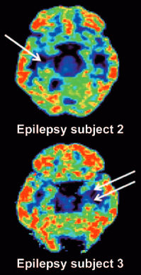
PET scan reveals unilateral mesial temporal sclerosis in epilepsy patients (white arrows indicate loss of [11C]UCB-J binding in the mesial temporal lobe). (credit: Sjoerd J. Finnema et al./Science Translational Medicine)

They used this new imaging technique on baboons and humans, then applied mathematical tools to quantify synaptic density, and confirmed that the new method served as a marker for synaptic density. The method revealed synaptic loss in three patients with epilepsy compared to healthy individuals.

With this noninvasive method, researchers may now be able to follow the progression of many brain disorders by measuring changes in synaptic density over time or assess how well pharmaceuticals slow the loss of neurons.

Professor of radiology and biomedical imaging Richard Carson and his team plan future studies involving PET imaging of synapses for a variety of brain disorders.

Published July 20 in Science Translational Medicine, the study was supported in part by the Swebilius Foundation, UCB Pharma, and the National Center for Advancing Translational Science, a component of the National Institutes of Health.


Abstract of Imaging synaptic density in the living human brain

Chemical synapses are the predominant neuron-to-neuron contact in the central nervous system. Presynaptic boutons of neurons contain hundreds of vesicles filled with neurotransmitters, the diffusible signaling chemicals. Changes in the number of synapses are associated with numerous brain disorders, including Alzheimer’s disease and epilepsy. However, all current approaches for measuring synaptic density in humans require brain tissue from autopsy or surgical resection. We report the use of the synaptic vesicle glycoprotein 2A (SV2A) radioligand [11C]UCB-J combined with positron emission tomography (PET) to quantify synaptic density in the living human brain. Validation studies in a baboon confirmed that SV2A is an alternative synaptic density marker to synaptophysin. First-in-human PET studies demonstrated that [11C]UCB-J had excellent imaging properties. Finally, we confirmed that PET imaging of SV2A was sensitive to synaptic loss in patients with temporal lobe epilepsy. Thus, [11C]UCB-J PET imaging is a promising approach for in vivo quantification of synaptic density with several potential applications in diagnosis and therapeutic monitoring of neurological and psychiatric disorders.

Distinct stages of thinking revealed by brain activity patterns

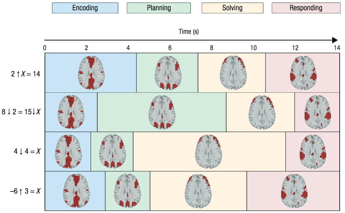
Durations of four stages associated with problem solving and corresponding MRI images. In the four example problems (left), the arrows denote new mathematical operators that participants had learned. Color coding reflects the durations of the stages. (credit: John R. Anderson et al./Psychological Science)

Using neuroimaging data, Carnegie Mellon University researchers have identified four distinct stages of math problem solving, according to a new study published in the journal Psychological Science.

“How students were solving these kinds of problems was a total mystery to us until we applied these techniques,” says psychological scientist John Anderson, lead researcher on the study. “Now, when students are sitting there thinking hard, we can tell what they are thinking each second.”

Insights from this work may eventually be applied to the design of more effective classroom instruction, says Anderson.

Combining pattern analysis and hidden semi-Markov models

Anderson combined two analytical approaches — multivoxel pattern analysis (MVPA) and hidden semi-Markov models (HSMM) — to shed light on the different stages of thinking. MVPA has typically been used to identify momentary patterns of activation; adding HSMM, Anderson hypothesized, would yield information about how these patterns play out over time.

The researchers applied this combined approach to neuroimaging data collected from participants as they solved specific types of math problems. To gauge whether the stages that were identified mapped on to actual stages of thinking, the researchers manipulated different features of the math problems; some problems required more effort in coming up with an appropriate solution plan and others required more effort in executing the solution.

The aim was to test whether these manipulations had the specific effects one would expect on the durations of the different stages.

Stages of cognition

The researchers identified four stages of cognition: encoding, planning, solving, and responding. The planning stage tended to be longer when the problem required more planning, and the solution stage tended to be longer when the solution was more difficult to execute, indicating that the method mapped onto real stages of cognition that were differentially affected by various features of the problems.

“Typically, researchers have looked at the total time to complete a task as evidence of the stages involved in performing that task and how they are related,” says Anderson.  “The methods in this paper allow us to measure the stages directly.”

Although the study focused specifically on mathematical problem solving, the method holds promise for broader application, the researchers argue. Using the same method with brain imaging techniques that have greater temporal resolution, such as EEG, could reveal even more detailed information about the various stages of cognitive processing.

This work was supported by a National Science Foundation grant and by a James S. McDonnell Foundation Scholar Award.


Abstract of Hidden Stages of Cognition Revealed in Patterns of Brain Activation

To advance cognitive theory, researchers must be able to parse the performance of a task into its significant mental stages. In this article, we describe a new method that uses functional MRI brain activation to identify when participants are engaged in different cognitive stages on individual trials. The method combines multivoxel pattern analysis to identify cognitive stages and hidden semi-Markov models to identify their durations. This method, applied to a problem-solving task, identified four distinct stages: encoding, planning, solving, and responding. We examined whether these stages corresponded to their ascribed functions by testing whether they are affected by appropriate factors. Planning-stage duration increased as the method for solving the problem became less obvious, whereas solving-stage duration increased as the number of calculations to produce the answer increased. Responding-stage duration increased with the difficulty of the motor actions required to produce the answer.

Mental, physical exercises found to produce different brain benefits

(credit: iStock)

Cognitive brain training improves executive function while aerobic activity improves memory, according to a new study by the Center for BrainHealth at The University of Texas at Dallas.

The study, published in an open-access paper in Frontiers in Human Neuroscience, compared cerebral blood flow and cerebrovascular reactivity data, obtained via MRI, for two groups of healthy sedentary adults ages 56–75 years. The members of both groups participated in training three hours per week over 12 weeks.

Cognitive training group

This group participated in cognitive training called Strategic Memory Advanced Reasoning Training (SMART), developed at the Center for BrainHealth. It focuses on three executive functions: strategic attention (prioritizing brain resources); integrative reasoning (synthesizing information at a deeper level); and innovation (encouraging fluid thinking, diverse perspective-taking, and problem solving).

The group demonstrated positive changes in executive brain function and a 7.9 percent increase in global brain flow.

“We can lose 1–2 percent in global brain blood flow every decade, starting in our 20s. To see almost an 8 percent increase in brain blood flow may be seen as regaining decades of brain health, since blood flow is linked to neural health,” said Sandra Bond Chapman, PhD, study lead author, founder and chief director of the Center for BrainHealth, and Dee Wyly Distinguished University Professor.

“We believe the reasoning training triggered neural plasticity by engaging the brain networks involved in staying focused on a goal, such as writing a brief business proposal, while continuously adapting to new information, such as feedback from a collaborator,” Chapman said.

Aerobic exercise group

The aerobic exercise group completed three, 60-minute sessions per week that included five minutes of warmup and cool down with 50 minutes of either walking on a treadmill or cycling on a stationary bike while maintaining 50–75 percent of maximum heart rate. It was designed to meet health guidelines for adults.

The group showed increases in immediate and delayed memory performance, with higher cerebral blood flow in the bilateral hippocampi, an area underlying memory function and particularly vulnerable to aging and dementia. But the group did not show significant global blood flow gains.

This work was supported by a grant from the National Institutes of Health and by grants from the Lyda Hill Foundation, T. Boone Pickens Foundation, and the Dee Wyly Distinguished University Endowment.


Abstract of Distinct Brain and Behavioral Benefits from Cognitive vs. Physical Training: A Randomized Trial in Aging Adults

Insidious declines in normal aging are well-established. Emerging evidence suggests that non-pharmacological interventions, specifically cognitive and physical training, may counter diminishing age-related cognitive and brain functions. This randomized trial compared effects of two training protocols: cognitive training (CT) vs. physical training (PT) on cognition and brain function in adults 56–75 years. Sedentary participants (N = 36) were randomized to either CT or PT group for 3 h/week over 12 weeks. They were assessed at baseline-, mid-, and post-training using neurocognitive, MRI, and physiological measures. The CT group improved on executive function whereas PT group’s memory was enhanced. Uniquely deploying cerebral blood flow (CBF) and cerebral vascular reactivity (CVR) MRI, the CT cohort showed increased CBF within the prefrontal and middle/posterior cingulate cortex (PCC) without change to CVR compared to PT group. Improvements in complex abstraction were positively associated with increased resting CBF in dorsal anterior cingulate cortex (dACC). Exercisers with higher CBF in hippocampi bilaterally showed better immediate memory. The preliminary evidence indicates that increased cognitive and physical activity improves brain health in distinct ways. Reasoning training enhanced frontal networks shown to be integral to top-down cognitive control and brain resilience. Evidence of increased resting CBF without changes to CVR implicates increased neural health rather than improved vascular response. Exercise did not improve cerebrovascular response, although CBF increased in hippocampi of those with memory gains. Distinct benefits incentivize testing effectiveness of combined protocols to strengthen brain health.

Facebook’s internet-beaming drone completes first test flight

(credit: Facebook)

Facebook Connectivity Lab announced today the first full-scale test flight of Aquila — a solar-powered unmanned airplane/drone designed to bring affordable internet access to some of the 1.6 billion people living in remote locations with no access to mobile broadband networks.

When complete, Aquila will be able to circle a region up to 60 miles in diameter, beaming internet connectivity down from an altitude of more than 60,000 feet to people within a 60-mile communications diameter for up to 90 days at a time. It will be part of a future fleet of drones.


Facebook

New nanomaterial mimics cell membranes

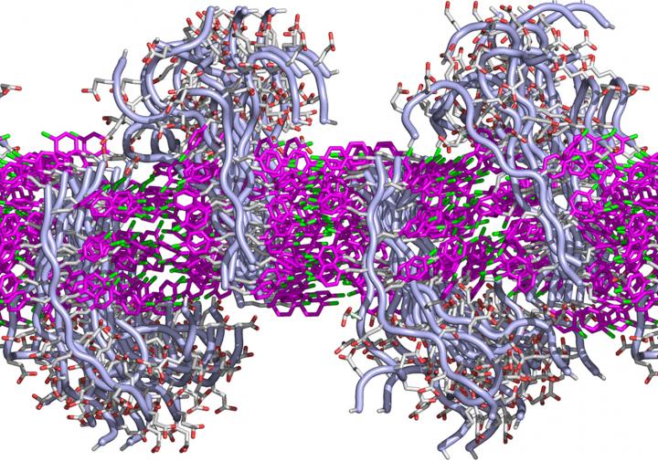
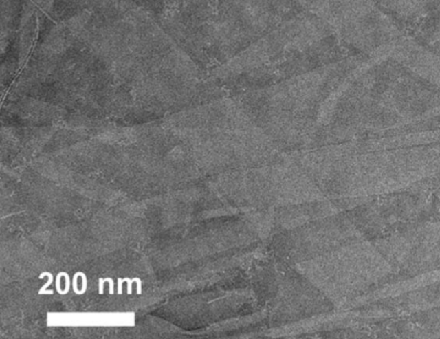
This simulated cross-section shows how the lipid-like peptoids interact to form a membrane. Each peptoid has two sections: a fatty-like region that interacts via benzene rings (shown in pink) with its neighbors to form a sheet, and a water-loving region that juts above or below the flat sheet. Each region can be designed to have specific functions. (credit: Chun-Long Chen/PNNL)

Materials scientists at the Department of Energy’s Pacific Northwest National Laboratory have created a new material that performs like a biological cell membrane — a material that has long been sought for applications like water purification and drug delivery.

The “peptoid” material can assemble itself into a sheet that’s thinner, but more stable, than a soap bubble, the researchers report this week in Nature Communications. The assembled sheet can withstand being submerged in a variety of liquids and can even repair itself after damage.

“We believe these materials have potential in water filters, sensors, drug delivery, and especially fuel cells or other energy applications,” said chemist Chun-Long Chen.

Biological cell membranes, which are made from thin sheets of fatty molecules called lipids, are at least ten times thinner than an iridescent soap bubble and yet allow cells to collectively form organisms as diverse at bacteria, trees and people.

Cell membranes are also very selective about what they let pass through, using tiny embedded proteins as gatekeepers. Membranes repair dings to their structure automatically and change thickness to pass signals from the outside environment to the cell’s interior, where most of the action is.

Scientists would like to take advantage of these gatekeeping and other membrane properties to make filters. A cell-membrane-like material would have advantages over other thin materials such as graphene. For example, mimicking a cell membrane’s efficient gatekeeping could result in water purifying membranes that don’t require a lot of pressure or energy to push the water through.

How to design imitation biological cell membranes

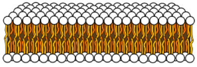
Lipid bilayer sheet (credit: Mariana Ruiz Villarreal/Wikipedia)

Synthetic molecules called peptoids have caught the interest of researchers because they are cheap, versatile and customizable. They are like natural proteins, including those that embed themselves in cell membranes, and can be designed to have very specific forms and functions. So Chen and colleagues decided to see if they could design peptoids to make them more lipid-like (that is, more like fats).

Lipid molecules are long and mostly straight: They have a fatty end that prefers to hang out with other fats, and a water-like end that prefers the comfort of water. Because of this chemistry, lipid molecules arrange themselves with the fatty ends pointed toward each other, sandwiched between the water-loving ends pointed out. Scientists call this a lipid bilayer, essentially a sheet that envelops the contents of a cell. Proteins or carbohydrate molecules embed themselves in the membranous sheet.

Inspired by this, Chen and colleagues designed peptoids in which each base peptoid was a long molecule with one end water-loving and the other end fat-loving. They chose chemical features that they hoped would encourage the individual molecules to pack together. They examined the resulting structures using a variety of analysis methods, including some at the Advanced Light Source and the Molecular Foundry, two DOE Office of Science User Facilities at Lawrence Berkeley National Laboratory.

Forming nanomembranes

Peptoid nanomembrane (credit: Haibao Jin et al./Nature Communications)

The team found that after putting the lipid-like peptoids into a liquid solution, the molecules spontaneously crystallized and formed “nanomembranes” — straight-edged sheets as thin as cell membranes — floating in the beaker. These nanomembranes maintained their structure in water or alcohol, at different temperatures, in solutions with high or low pH, or high concentrations of salts, a feat that few cell membranes could accomplish.

To better understand the nanomembranes, the team simulated how single peptoid molecules interacted with each other using molecular dynamics software. The simulated peptoids formed a membrane reminiscent of a lipid bilayer: The fat-loving ends lined up in the middle, and their water-loving ends pointed outward either above or below.

They also confirmed the ability of the synthetic membranes to hold proteins that have specific functions, such as ones that let water, and only water, through, and to repair themselves.

The results showed the researchers that they are on the right path to making synthetic cell membrane-like materials. The next step, Chen said, is to build biomimetic membranes by incorporating natural membrane proteins or other synthetic water channels such as carbon nanotubes into these sheet matrices. The team is also looking into ways to make the peptoid membranes conductive for energy uses.

This work was supported by the Department of Energy Office of Science and PNNL.


Abstract of Highly stable and self-repairing membrane-mimetic 2D nanomaterials assembled from lipid-like peptoids

An ability to develop sequence-defined synthetic polymers that both mimic lipid amphiphilicity for self-assembly of highly stable membrane-mimetic 2D nanomaterials and exhibit protein-like functionality would revolutionize the development of biomimetic membranes. Here we report the assembly of lipid-like peptoids into highly stable, crystalline, free-standing and self-repairing membrane-mimetic 2D nanomaterials through a facile crystallization process. Both experimental and molecular dynamics simulation results show that peptoids assemble into membranes through an anisotropic formation process. We further demonstrated the use of peptoid membranes as a robust platform to incorporate and pattern functional objects through large side-chain diversity and/or co-crystallization approaches. Similar to lipid membranes, peptoid membranes exhibit changes in thickness upon exposure to external stimuli; they can coat surfaces in single layers and self-repair. We anticipate that this new class of membrane-mimetic 2D nanomaterials will provide a robust matrix for development of biomimetic membranes tailored to specific applications.

Musk’s new master plan for Tesla

Tesla Autopilot (credit: Tesla Motors)

Elon Musk revealed his new master plan for Tesla today (July 20) in a blog post published on Tesla’s website:

  • Create stunning solar roofs with seamlessly integrated battery storage.
  • Expand the electric vehicle product line to address all major segments.
  • Develop a self-driving capability that is 10X safer than manual via massive fleet learning.
  • Enable your car to make money for you when you aren’t using it.

Increasing safety: “morally reprehensible to delay”

In the context of the recent Autopilot problem, Musk clarified why Tesla is deploying partial autonomy now, rather than waiting until some point in the future: “When used correctly, it is already significantly safer than a person driving by themselves and it would therefore be morally reprehensible to delay release simply for fear of bad press or some mercantile calculation of legal liability.

“According to the recently released 2015 NHTSA report, automotive fatalities increased by 8% to one death every 89 million miles. Autopilot miles will soon exceed twice that number and the system gets better every day. It would no more make sense to disable Tesla’s Autopilot, as some have called for, than it would to disable autopilot in aircraft, after which our system is named.”

Another way to increase safety, he says, is new heavy-duty trucks and high passenger-density urban transport, both planned for unveiling next year. “With the advent of autonomy, it will probably make sense to shrink the size of buses and transition the role of bus driver to that of fleet manager. … Traffic congestion would improve due to increased passenger areal density by eliminating the center aisle and putting seats where there are currently entryways, and matching acceleration and braking to other vehicles, thus avoiding the inertial impedance to smooth traffic flow of traditional heavy buses. It would also take people all the way to their destination.”

Lowering the cost of an autonomous car

Musk said that when true self-driving is approved by regulators, “it will mean that you will be able to summon your Tesla from pretty much anywhere. Once it picks you up, you will be able to sleep, read, or do anything else enroute to your destination.

“You will also be able to add your car to the Tesla shared fleet just by tapping a button on the Tesla phone app and have it generate income for you while you’re at work or on vacation, significantly offsetting and at times potentially exceeding the monthly loan or lease cost. This dramatically lowers the true cost of ownership to the point where almost anyone could own a Tesla. Since most cars are only in use by their owner for 5% to 10% of the day, the fundamental economic utility of a true self-driving car is likely to be several times that of a car which is not.”

Musk said that in cities where demand exceeds the supply of customer-owned cars, “Tesla will operate its own fleet, ensuring you can always hail a ride from us no matter where you are.”

Why partially automated cars should be deployed in ‘light-duty vehicles’

Crash avoidance technologies now available in non-luxury vehicles include Lane Departure Warning (LDW), Forward Collision Warning (FCW), and Blind Spot Monitoring Roadway (BSM). (credit: Corey D. Harper et al./Accident Analysis and Prevention)

U.S. National Highway Traffic Safety Administration chief Mark Rosekind said at a conference today (July 20) that the government “will not abandon efforts to speed the development of self-driving cars … to reduce the 94 percent of car crashes attributed to human error, despite a fatal accident involving a Tesla Model S operating on an autopilot system,” Reuters reports. But autonomous vehicles must be “much safer” than human drivers before they are deployed on U.S. roads, he added.

However, Carnegie Mellon College of Engineering researchers suggest that already-available partially automated vehicle crash-avoidance technologies are a practical interim solution, they conclude in a study published in the journal Accident Analysis and Prevention.

These technologies — which include forward collision warning and avoidance, lane departure warning, blind spot monitoring, and partially autonomous braking or controls — are already available in non-luxury vehicles such as the Honda Accord and Mazda CX-9. If these technologies were deployed in all “light-duty vehicles,” it could prevent or reduce the severity of up 1.3 million crashes a year, including 10,100 fatal wrecks, according to the study.

“While there is much discussion about driverless vehicles, we have demonstrated that even with partial automation, there are financial and safety benefits,” says Chris T. Hendrickson, director of the Carnegie Mellon Traffic21 Institute.

When the team compared the price of equipping cars with safety technology to the expected annual reduction in the costs of crashes (based on government and insurance industry data), they discovered a net cost benefit (in addition to life-saving benefits) in two scenarios:

  • In the perfect-world scenario in which all relevant crashes are avoided with these technologies, there is an annual benefit of $202 billion or $861 per car.
  • On the more conservative side, when only observed crash reductions in vehicles equipped with blind spot monitoring, lane departure and forward collision crash avoidance systems are considered, there is still an annual positive net benefit of $4 billion dollars or $20 a vehicle (and lower prices could lead to larger net benefits over time).

Carnegie Mellon’s Technologies for Safe and Efficient Transportation (T-SET) University Transportation Center, the National Science Foundation, and the Hillman Foundation funded the project.


Abstract of Cost and benefit estimates of partially-automated vehicle collision avoidance technologies

Many light-duty vehicle crashes occur due to human error and distracted driving. Partially-automated crash avoidance features offer the potential to reduce the frequency and severity of vehicle crashes that occur due to distracted driving and/or human error by assisting in maintaining control of the vehicle or issuing alerts if a potentially dangerous situation is detected. This paper evaluates the benefits and costs of fleet-wide deployment of blind spot monitoring, lane departure warning, and forward collision warning crash avoidance systems within the US light-duty vehicle fleet. The three crash avoidance technologies could collectively prevent or reduce the severity of as many as 1.3 million U.S. crashes a year including 133,000 injury crashes and 10,100 fatal crashes. For this paper we made two estimates of potential benefits in the United States: (1) the upper bound fleet-wide technology diffusion benefits by assuming all relevant crashes are avoided and (2) the lower bound fleet-wide benefits of the three technologies based on observed insurance data. The latter represents a lower bound as technology is improved over time and cost reduced with scale economies and technology improvement. All three technologies could collectively provide a lower bound annual benefit of about $18 billion if equipped on all light-duty vehicles. With 2015 pricing of safety options, the total annual costs to equip all light-duty vehicles with the three technologies would be about $13 billion, resulting in an annual net benefit of about $4 billion or a $20 per vehicle net benefit. By assuming all relevant crashes are avoided, the total upper bound annual net benefit from all three technologies combined is about $202 billion or an $861 per vehicle net benefit, at current technology costs. The technologies we are exploring in this paper represent an early form of vehicle automation and a positive net benefit suggests the fleet-wide adoption of these technologies would be beneficial from an economic and social perspective.

Peering into atomically thin transistors with microwaves, scientists make a radical discovery: a one-dimensional transistor

Peering into an atomically thin semiconductor device with a “microwave microscope” via an AFM tip (credit: University of Texas at Austin)

Physicists at The University of Texas at Austin have had the first-ever glimpse into what happens inside an atomically thin semiconductor device. In doing so, they discovered that a transistor may be possible within a space so small (the edge) that it’s effectively one-dimensional.

Future tech innovations will require finding a way to fit more transistors on computer chips to keep up with Moore’s law, so experts have begun exploring new semiconducting materials, including one called molybdenum disulfide (MoS2), as KurzweilAI reported last week. In a paper published July 18 in the Proceedings of the National Academy of Sciences, the researchers describe seeing the detailed inner workings of this new type of two-dimensional transistor.

Unlike today’s silicon-based devices, transistors made from MoS2 allow for on-off signaling on a single 2-D (flat) plane.

Computing on a one-dimensional edge

Keji Lai*, an assistant professor of physics, and his team used a “microwave impedance microscope” that he invented and that pointed microwaves at the 2-D device. Using an atomic force microscope (AFM) tip only 100 nanometers wide, the microwave microscope allowed the scientists to see conductivity changes inside the transistor for the first time.

That’s when they discovered that with MoS2, the conductive signaling happens much differently than with silicon — in a way that could promote future energy savings in devices.

With silicon transistors, the entire device is either turned on or off. With 2-D transistors, by contrast, Lai and the team found that electric currents move in a more phased (or wave-like) way, beginning first at the edges before appearing in the interior. Lai says this suggests the same current could be sent with less power and in an even tinier space — using a one-dimensional edge (a line) instead of the two-dimensional plane (area).

“In physics, edge states often carry a lot of interesting phenomena, and here, they are the first to turn on. In the future, if we can engineer this material very carefully, then these edges can carry the full current,” Lai says. “We don’t really need the entire thing, because the interior is useless. Just having the edges running to get a current working would substantially reduce the power loss.”

Eliminating defects

Researchers have been working to get a view into what happens inside a 2-D transistor for years to better understand both the potential and the limitations of the new materials. Getting 2-D transistors ready for commercial devices, such as paper-thin computers and cellphones, is expected to take several more years. Lai says scientists need more information about what interferes with performance in devices made from the new materials.

Besides seeing the currents’ motion, the scientists found thread-like defects in the middle of the transistors. Lai says this suggests the new material will need to be made cleaner to function optimally. “If we could make the material clean enough, the edges will be carrying even more current, and the interior won’t have as many defects,” Lai says.

The research was supported by the U.S. Department of Energy, the Welch Foundation, the Office of Naval Research, and the National Science Foundation.

* Earlier this year, Lai and co-researcher Deji Akinwande, associate professor of UT Austin’s Department of Electrical and Computer Engineering, won Presidential Early Career Awards for Scientists and Engineers, the U.S. government’s highest honor for early-stage scientists and engineers.


UT | In this visualization of what happens inside a 2-D transistor made of a promising new material called MoS2, electric currents appear initially at the outer edges and then inside of the device. Thread-like flaws can be seen in the interior part of the transistor.


Abstract of Uncovering edge states and electrical inhomogeneity in MoS2 field-effect transistors

The understanding of various types of disorders in atomically thin transition metal dichalcogenides (TMDs), including dangling bonds at the edges, chalcogen deficiencies in the bulk, and charges in the substrate, is of fundamental importance for TMD applications in electronics and photonics. Because of the imperfections, electrons moving on these 2D crystals experience a spatially nonuniform Coulomb environment, whose effect on the charge transport has not been microscopically studied. Here, we report the mesoscopic conductance mapping in monolayer and few-layer MoS2 field-effect transistors by microwave impedance microscopy (MIM). The spatial evolution of the insulator-to-metal transition is clearly resolved. Interestingly, as the transistors are gradually turned on, electrical conduction emerges initially at the edges before appearing in the bulk of MoS2 flakes, which can be explained by our firstprinciples calculations. The results unambiguously confirm that the contribution of edge states to the channel conductance is significant under the threshold voltage but negligible once the bulk of the TMD device becomes conductive. Strong conductance inhomogeneity, which is associated with the fluctuations of disorder potential in the 2D sheets, is also observed in the MIM images, providing a guideline for future improvement of the device performance.

New brain map provides unprecedented detail in 180 areas of the cerebral cortex

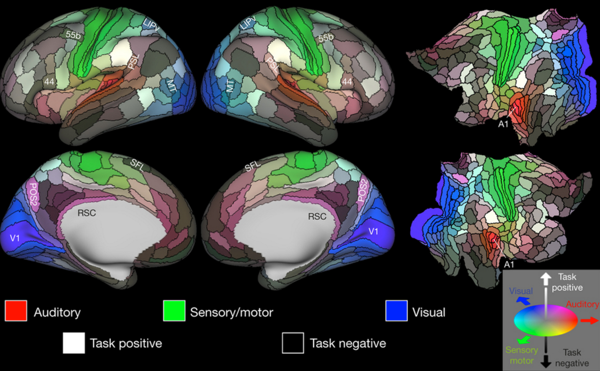
A detailed new map by researchers at Washington University School of Medicine in St. Louis lays out the landscape of the cerebral cortex. The 180 areas delineated and identified in both left and right hemispheres are displayed on inflated and flattened cortical surfaces. Black outlines indicate areal borders. Colors indicate the extent to which the areas are associated in the resting state with auditory (red), somatosensory (green), visual (blue), task positive (towards white), or task negative (towards black) groups of areas. The legend on the bottom right illustrates the 3D color space used in the figure. Data at http://balsa.wustl.edu/WN56. (credit: Matthew F. Glasser et al./Nature)

A detailed new map by researchers at Washington University School of Medicine in St. Louis and associates* lays out the landscape of 180 areas of the cerebral cortex in painstaking detail; 97 of these areas have never been previously described.

The new map is intended to help researchers studying brain disorders such as autism, schizophrenia, dementia and epilepsy. They will be able to use it to understand differences in the brains of patients with these diseases, compared with adults who are healthy. It also will accelerate progress in deciphering the workings of the healthy brain and elucidating what makes us unique as a species, the researchers say. The new map will also be vital for neurosurgeons.

The work was published today, July 20, in Nature.


Nature Video | The ultimate brain map

The researchers drew upon data and methods generated by the Human Connectome Project, a five-year, multimillion dollar study led by David Van Essen, PhD, the senior author on this paper. The Human Connectome Project used a powerful, custom-built MRI machine to map the brains of 1,200 young adults. This new study complements the Human Connectome Project by carefully delineating the brain regions so that their connections can be more accurately mapped.

The new map divides both the left and right cerebral hemispheres into 180 areas based on physical differences (such as the thickness of the cortex), functional distinctions (such as which areas respond to language stimuli), and differences in the connections of the areas.

“The brain is not like a computer that can support any operating system and run any software,” said Van Essen, the Alumni Endowed Professor of Neuroscience at Washington University Medical School. “Instead, the software — how the brain works — is intimately correlated with the brain’s structure, its hardware, so to speak. If you want to find out what the brain can do, you have to understand how it is organized and wired.”

The researchers mapped the cortex, a layer of neural tissue that encases the rest of the brain like a crumpled sheet of paper. The cortex is important for sensation, attention, memory, perception, thought, language and consciousness.

Beyond Brodmann’s areas

Brodmann areas (credit: Mark Dow/Wikipedia)

The new study is intended to replace previous maps, such as Korbinian Brodmann’s map of the human cortex, created in the first decade of the 20th century, which identified 50 regions. “My early work on language connectivity involved taking that 100-year-old map and trying to guess where Brodmann’s areas were in relation to the pathways underneath them,” said Matthew Glasser, PhD, of Washington University Medical School. “It quickly became obvious to me that we needed a better way to map the areas in the living brains that we were studying.”

Until now, most brain maps have been based on a single type of measurement. To make the new map, the researchers pooled data from 210 healthy young adults of both sexes, combining measures of the thickness of the cortex and the amount of insulation around neuronal cables, using MRI scans of the resting brain and of the brain performing simple tasks, such as listening to a story. The information also included measurements of brain function, connectivity between regions, topographic organization of cells in brain tissue, and levels of myelin, which speeds up neural signaling.

In the new map, some of the 180 areas identified are clearly involved in particular tasks, such as 55b, which lights up with activity when a person hears a story. Others contain a map of a person’s field of vision, or are involved in controlling movement. But most areas probably will never be identified with a single function, because they don’t do just one thing, but instead coordinate information from many different signals.

Many other maps of the cortex have been drawn, showing anywhere from 50 to 200 different areas. But the researchers say they improved on previous maps by precisely aligning the brains to a common coordinate system before analysis, using an algorithm developed by colleagues at Oxford University, and incorporating the highest-quality MRI data available. The researchers also verified that their method could be applied to individuals by producing maps of the brains of a different set of 210 healthy young adults.

Guiding neurosurgeons

The results are a precise map with unusually crisp borders and an algorithm capable of locating the areas in individual brains, even though each individual is unique in terms of the pattern of cortical folds and in the size and shape of areas on the cortical map.

“In the past, it was not always clear whether the results from two separate neuroimaging studies referred to the same area or not,” Glasser said. By using the new map and alignment algorithm, results of separate studies could be more accurately compared.

Better individual maps of the brain could help neurosurgeons avoid damaging the most important areas, such as those involved in language or motor function, and could guide treatment for neurological or psychiatric illnesses. Different types of dementia, for example, are characterized by degeneration in different areas of the brain. Clinicians could use the individual maps to personalize treatment, based on the areas affected, or to monitor response to treatment.

Like cartographers of old, brain cartographers primarily are providing a tool for others to use in exploration and discovery.

“We were able to persuade Nature to put online almost 200 extra pages of detailed information on each of the 180 regions as well as all of the algorithms we used to align the brains and create the map,” Van Essen said. “We think it will serve the scientific community best if they can dive down and get these maps onto their computer screens and explore as they see fit.”

* University of Oxford; Imperial College, London; University of Minnesota; Radboud University; Radboud University Medical Centre.