US has potential to produce more than a billion tons of biomass annually by 2040


Oak Ridge National Laboratory | 2016 Billion-Ton Report

The U.S. has the potential to sustainably produce at least 1 billion dry tons of nonfood biomass resources annually by 2040, according to the 2016 Billion-Ton Report, jointly released by the U.S. Department of Energy and Oak Ridge National Laboratory. That amount would substantially decrease greenhouse gas emissions in the utility and transportation sectors and (as the domestic bioeconomy grows) reduce U.S. dependence on imported oil, the scientists project.

These renewable resources include agricultural, forestry and algal biomass, as well as waste. They encompass the current and future potential of biomass, from currently available logging and crop residues to future available algae and dedicated energy crops — all useable for the production of biofuel, biopower and bioproducts.

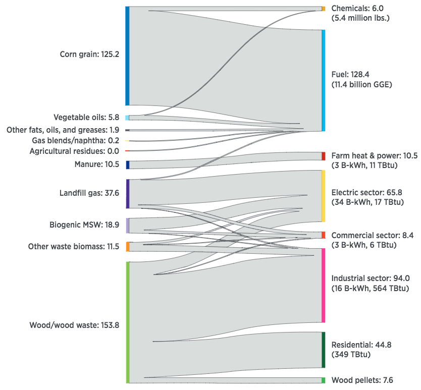
Current feedstock, sector consumption, and final product distribution, in million dry tons per year. Biomass resources are shown on the left and their allocations are shown on the right. The size of the flow is representative of the amount of biomass allocated to that end use. For this figure, contributions from landfill gas are represented as tons of biomass equivalent by applying a conversion factor of 0.2665 lb/scf (credit: U.S. DOE)

The report findings show that under a base-case scenario, the United States could increase its use of dry biomass resources from a current 400 million tons to 1.57 billion tons under a high-yield scenario.

The analysis was led by ORNL with contributions from 65 experts from federal agencies, national laboratories, universities (the University of Tennessee, North Carolina State University, South Dakota State University and Oregon State University), and private companies (Energetics, Inc. and Allegheny Science and Technology).

Proposed future feedstock supply system for transforming raw biomass into stable, tradeable
commodities suitable for long-distance transport and handling in existing infrastructure (credit: Idaho National Laboratory)

New to the 2016 report are assessments of potential biomass supplies from algae, from new energy crops (miscanthus, energy cane, eucalyptus), and from municipal solid waste. For the first time, the report also considers how the cost of pre-processing and transporting biomass to the biorefinery may impact feedstock availability.

Interactive tools available through the Bioenergy Knowledge Discovery Framework allow users to visualize biomass availability scenarios and  tailor the data by factors such as geographic area, biomass source and price. Researchers and decision makers can use these features to better inform national bioenergy policies and research, development and deployment strategies. Each diagram and map in the report is available in an interactive interface on the Bioenergy Knowledge Discovery Framework.

Volume 2 of the report, set for release later this year, will consist of a collection of analyses on the potential environmental sustainability effects of a subset of agricultural and forestry biomass production scenarios presented in volume 1. Volume 2 will also discuss algae sustainability, land use, and land management changes, and strategies to enhance environmental sustainability.

World’s smallest storage device writes information atom by atom

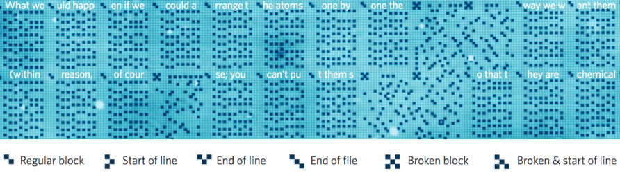
STM scan (96 nm wide, 126 nm tall) of the 1 kB memory, written to a section of Feynman’s lecture, “There’s Plenty of Room at the Bottom” (credit: TU Delft/Ottelab)

Scientists at Kavli Institute of Nanoscience at Delft University have built a nanoscale data storage device containing 1 kilobyte (8,000 bits) with a storage density of 500 terabits per square inch (Tbpsi) — 500 times denser than the best commercial hard disk drive currently available. Each bit is represented by the position of one single chlorine atom.

“In theory, this storage density would allow all books ever created by humans to be written on a single post stamp,” says lead scientist Sander Otte. The research is reported today (Monday July 18) in Nature Nanotechnology.

Every day, modern society creates more than a billion gigabytes of new data. To store all this data, it is increasingly important that each single bit occupies as little space as possible.

In 1959, physicist Richard Feynman challenged his colleagues to engineer the world at the smallest possible scale. In his famous lecture There’s Plenty of Room at the Bottom, he speculated that if we had a platform allowing us to arrange individual atoms in an exact orderly pattern, it would be possible to store one piece of information per atom. To honor the visionary Feynman, Otte and his team have coded a section of Feynman’s lecture on an area 100 nanometers wide.

“Sliding puzzle” scheme

Atomic data storage scheme (credit: Kavli Institute of Nanoscience)

The team used a scanning tunneling microscope (STM), in which a sharp needle probes the atoms of a surface, one by one. With these probes scientists can see atoms and push them around. “You could compare it  to a sliding puzzle,” Otte explains. “Every bit consists of two positions on a surface of copper atoms, and one chlorine atom that we can slide back and forth between these two positions. If the chlorine atom is in the top position, there is a hole beneath it — we call this a 1. If the hole is in the top position and the chlorine atom is therefore on the bottom, then the bit is a 0.”

Because the chlorine atoms are surrounded by other chlorine atoms, except near the holes, they keep each other in place. Which is why this method with holes is much more stable than methods with loose atoms and more suitable for data storage.

Kilobyte atomic memory. 1,016-byte atomic memory, written to a passage from Feynman’s lecture, “There’s plenty of room at the bottom.” The memory consists of 127 functional blocks and 17 broken blocks, resulting in an overall areal density of 0.778 bits per nm square. (credit: F. E. Kalff et al./Nature Nanotechnology)

The researchers organized their memory in blocks of 8 bytes (64 bits). Each block has a marker, made of the same type of “holes” as the raster of chlorine atoms. Inspired by the pixelated square barcodes (QR codes) often used to scan tickets for airplanes and concerts, these markers work like miniature QR codes that carry information about the precise location of the block on the copper layer. The code will also indicate if a block is damaged, for instance due to some local contaminant or an error in the surface. This allows the memory to be scaled up easily to very big sizes, even if the copper surface is not entirely perfect.

The new approach offers excellent prospects in terms of stability and scalability. However, “in its current form the memory can operate only in very clean vacuum conditions and at liquid nitrogen temperature (77 K), so the actual storage of data on an atomic scale is still some way off.”

This research was support by the Netherlands Organisation for Scientific Research (NOW/FOM). Scientists of the International Iberian Nanotechnology Laboratory (INL) in Portugal performed calculations on the behavior of the chlorine atoms.


Delft University of Technology | Atomic scale data storage


Abstract of A kilobyte rewritable atomic memory

The advent of devices based on single dopants, such as the single-atom transistor, the single-spin magnetometer and the single-atom memory, has motivated the quest for strategies that permit the control of matter with atomic precision. Manipulation of individual atoms by low-temperature scanning tunnelling microscopy provides ways to store data in atoms, encoded either into their charge state, magnetization state or lattice position. A clear challenge now is the controlled integration of these individual functional atoms into extended, scalable atomic circuits. Here, we present a robust digital atomic-scale memory of up to 1 kilobyte (8,000 bits) using an array of individual surface vacancies in a chlorine-terminated Cu(100) surface. The memory can be read and rewritten automatically by means of atomic-scale markers and offers an areal density of 502 terabits per square inch, outperforming state-of-the-art hard disk drives by three orders of magnitude. Furthermore, the chlorine vacancies are found to be stable at temperatures up to 77 K, offering the potential for expanding large-scale atomic assembly towards ambient conditions.

Middle-age-plus memory decline may just be a matter of changing focus

When middle-aged and older adults were shown a series of faces, red regions of the brain were more active; these include an area in the medial prefrontal cortex that is associated with self-referential thinking. In young adults, by contrast, blue regions — which include areas important for memory and attention –+ were more active during this task. (credit: N. Rajah, McGill University)

Are you middle-aged or older and having problems remembering details, like where you left the keys or parked your car?

Cheer up, it may simply be the result of a change in what information your brain focuses on during memory formation and retrieval, rather than a decline in brain function, according to a study by McGill University researchers.

In the study, published in the journal, NeuroImage, 112 healthy adults ranging in age from 19 to 76 years were shown a series of faces. Participants were then asked to recall where a particular face appeared on the screen (left or right) and when it appeared (least or most recently). The researchers used functional MRI to analyze which parts of brain were activated during recall of these details.

Different parts of the brain involved

Senior author Natasha Rajah, Director of the Brain Imaging Centre, and colleagues found that young adults activated their visual cortex while successfully performing this task.

But for middle-aged and older adults, their medial prefrontal cortex was activated instead. That’s a part of the brain known to be involved with information having to do with one’s own life and introspection. This may reflect changes in what adults deem “important information” as they age, she said.

Rajah says middle-aged and older adults can improve their recall abilities by learning to focus on external rather than internal information, using mindfulness meditation, for example.*

Rajah is currently analyzing data from a similar study to discern if there are any gender differences in middle-aged brain function as it relates to memory. “At mid-life women are going through a lot of hormonal change. So we’re wondering how much of these results is driven by post-menopausal women.”

The research was supported by the Canadian Institutes of Health Research and by a grant from the Alzheimer’s Society of Canada.

* Other options to improve memory include:

  • Peppermint tea or rosemary essential oil, scientists at Northumbria University found in studies with subjects over age 65, presented at the British Psychological Society’s Annual Conference in Nottingham in April. Rosemary aroma significantly enhanced prospective memory (for things you plan to do).
  • Eight nutrients to protect the aging brain: cocoa flavanols, omega-3 fatty acids, phosphatidylserine and phosphatidic Acid, walnuts, citicoline, choline (found especially in eggs) and magnesium (avocado, soy beans, bananas and dark chocolate), and blueberries, according to a study published in the journal Food Technology. Details here.

Abstract of Changes in the modulation of brain activity during context encoding vs. context retrieval across the adult lifespan

Age-related deficits in context memory may arise from neural changes underlying both encoding and retrieval of context information. Although age-related functional changes in the brain regions supporting context memory begin at midlife, little is known about the functional changes with age that support context memory encoding and retrieval across the adult lifespan. We investigated how age-related functional changes support context memory across the adult lifespan by assessing linear changes with age during successful context encoding and retrieval. Using functional magnetic resonance imaging (fMRI), we compared young, middle-aged and older adults during both encoding and retrieval of spatial and temporal details of faces. Multivariate behavioral partial least squares (B-PLS) analysis of fMRI data identified a pattern of whole brain activity that correlated with a linear age term, and a pattern of whole brain activity that was associated with an age-by-memory phase (encoding vs. retrieval) interaction. Further investigation of this latter effect identified three main findings: 1) reduced phase-related modulation in bilateral fusiform gyrus, left superior/anterior frontal gyrus and right inferior frontal gyrus that started at midlife and continued to older age, 2) reduced phase-related modulation in bilateral inferior parietal lobule that occurred only in older age, and 3) changes in phase-related modulation in older but not younger adults in left middle frontal gyrus and bilateral parahippocampal gyrus that was indicative of age-related over-recruitment. We conclude that age-related reductions in context memory arise in midlife and are related to changes in perceptual recollection and changes in fronto-parietal retrieval monitoring.

Mayo Clinic researchers discover drug combination that helps immune system attack cancer cells

Effects of combination drug treatment on tumor size in square millimeters over 67 days for multiple mice (credit: Soraya Zorro Manrique et al./Oncotarget)

Mayo Clinic researchers have developed a drug combination that could enhance the immune system’s ability to attack cancer cells. The drugs have shown a pronounced therapeutic effect against advanced and metastatic cancers in mice, according to a  study published in the July 12 edition of the online journal Oncotarget.

“Cancers can remain inconspicuous in the body for months to years before causing major problems, leading the immune system to coexist rather than to attack cancers,” explains Mayo Clinic cancer immunotherapist Peter Cohen, M.D.

The solution was to combine two drugs: toll-like receptor (TLR) agonists that can mimic invading bacteria (tricking the immune system into attacking cancer as if it were a life-threatening infection) and the chemotherapy agent cyclophosphamide. The combination resulted in permanent eradication of breast and pancreatic cancers, as well as widespread metastases.

The combined weekly treatment was very well tolerated and actually less toxic than either TLR agonists or cyclophosphamide given individually, they also found.

The drug combination also revealed an additional benefit: it activated monocytes (a type of white blood cell) to participate in the killing of cancer cells.

Mayo Clinic is continuing its research in an FDA-approved clinical trial. They are studying whether patients with advanced cancers, including pancreas, breast, colorectal, melanoma and others, respond similarly to mice when cyclophosphamide treatment is paired with the TLR agonist motolimod.


Mayo Clinic | Potential Immunotherapy Drug Combination for Targeting Advanced and Metastatic Cancers


Abstract of Definitive activation of endogenous antitumor immunity by repetitive cycles of cyclophosphamide with interspersed Toll-like receptor agonists

Many cancers both evoke and subvert endogenous anti-tumor immunity. However, immunosuppression can be therapeutically reversed in subsets of cancer patients by treatments such as checkpoint inhibitors or Toll-like receptor agonists (TLRa). Moreover, chemotherapy can leukodeplete immunosuppressive host elements, including myeloid-derived suppressor cells (MDSCs) and regulatory T-cells (Tregs). We hypothesized that chemotherapy-induced leukodepletion could be immunopotentiated by co-administering TLRa to emulate a life-threatening infection. Combining CpG (ODN 1826) or CpG+poly(I:C) with cyclophosphamide (CY) resulted in uniquely well-tolerated therapeutic synergy, permanently eradicating advanced mouse tumors including 4T1 (breast), Panc02 (pancreas) and CT26 (colorectal). Definitive treatment required endogenous CD8+ and CD4+ IFNγ-producing T-cells. Tumor-specific IFNγ-producing T-cells persisted during CY-induced leukopenia, whereas Tregs were progressively eliminated, especially intratumorally. Spleen-associated MDSCs were cyclically depleted by CY+TLRa treatment, with residual monocytic MDSCs requiring only continued exposure to CpG or CpG+IFNγ to effectively attack malignant cells while sparing non-transformed cells. Such tumor destruction occurred despite upregulated tumor expression of Programmed Death Ligand-1, but could be blocked by clodronate-loaded liposomes to deplete phagocytic cells or by nitric oxide synthase inhibitors. CY+TLRa also induced tumoricidal myeloid cells in naive mice, indicating that CY+TLRa’s immunomodulatory impacts occurred in the complete absence of tumor-bearing, and that tumor-induced MDSCs were not an essential source of tumoricidal myeloid precursors. Repetitive CY+TLRa can therefore modulate endogenous immunity to eradicate advanced tumors without vaccinations or adoptive T-cell therapy. Human blood monocytes could be rendered similarly tumoricidal during in vitro activation with TLRa+IFNγ, underscoring the potential therapeutic relevance of these mouse tumor studies to cancer patients.

Why your immune system may control your social behavior

Normal brain activity (credit: University of Virginia Health System)

In a discovery that raises fundamental questions about human behavior, researchers at the University of Virginia School of Medicine have found that the immune system directly affects — and even controls — our social behavior, such as our desire to interact with others. That finding could have significant implications for neurological diseases such as autism-spectrum disorders and schizophrenia, the researchers suggest.

“The brain and the adaptive immune system were thought to be isolated from each other, and any immune activity in the brain was perceived as sign of a pathology. And now, not only are we showing that they are closely interacting, but some of our behavior traits might have evolved because of our immune response to pathogens,” explained Jonathan Kipnis, chair of UVA’s Department of Neuroscience.

“It’s crazy, but maybe we are just multicellular battlefields for two ancient forces: pathogens and the immune system. Part of our personality may actually be dictated by the immune system.”

Evolutionary forces linking brains and pathogens

KurzweilAI has cited supporting evidence for that idea. For example, permanent stress may affect immune cells in the brain, leading to mental disorders and protective immune microglia cells also have direct involvement in creating the cellular networks at the core of brain behavior.

Last year, Kipnis, the director of UVA’s Center for Brain Immunology and Glia, and his team discovered that meningeal membranes (covering the brain and spinal cord) in the brain directly link the brain with the lymphatic system. That overturned decades of textbook teaching that the brain lacks a direct connection to the immune system.

Now, the researchers suggest, the relationship between people and pathogens could have directly affected the development of our social behavior. Social behavior (which is necessary for the survival of the species) allows pathogens to spread, so our immune systems may have developed to protect us from the diseases that accompany those interactions.

Specifically, the UVA researchers have now shown that a specific immune molecule, interferon gamma, seems to be critical for social behavior, and that a variety of creatures, such as flies, zebrafish, mice and rats, activate interferon gamma responses (as protection) when they are social.

Normally, this molecule is produced by the immune system in response to bacteria, viruses or parasites. But blocking the molecule in mice using genetic modification also made regions of the brain hyperactive, causing the mice to become less social. Restoring the molecule restored the brain connectivity and behavior to normal.

A hyperactive brain, triggered by a blocked immune system, may lead to less-social behavior (credit: University of Virginia Health System)

In a Nature paper outlining their findings, the researchers note the immune molecule plays a “profound role in maintaining proper social function.”

“It’s extremely critical for an organism to be social for the survival of the species. It’s important for foraging, sexual reproduction, gathering, hunting,” said Anthony J. Filiano, Hartwell postdoctoral fellow in the Kipnis lab and lead author of the study. “The hypothesis is that when organisms come together, you have a higher propensity to spread infection — you need to be social, but [in doing so] you have a higher chance of spreading pathogens.” Which explains why “interferon gamma, in evolution, has been used as a more efficient way to both boost social behavior while boosting an anti-pathogen response.”

Immune-system failure leads to social deficits

The researchers explain that a malfunctioning immune system may be responsible for “social deficits in numerous neurological and psychiatric disorders.” But exactly what this might mean for autism and other specific conditions requires further investigation.

It is unlikely that any one molecule will be responsible for disease or the key to a cure. The researchers believe that the causes are likely to be much more complex. But the discovery that the immune system — and possibly pathogens, by extension — can control our interactions raises many exciting avenues for scientists to explore, both in terms of battling neurological disorders and understanding human behavior.

Kipnis and his team worked closely with UVA’s Department of Pharmacology and with Vladimir Litvak’s research group at the University of Massachusetts Medical School. Litvak’s team developed a computational approach to investigate the complex dialogue between immune signaling and brain function in health and disease.

“Using this approach we predicted a role for interferon gamma, an important cytokine secreted by T lymphocytes, in promoting social brain functions,” Litvak said. “Our findings contribute to a deeper understanding of social dysfunction in neurological disorders, such as autism and schizophrenia, and may open new avenues for therapeutic approaches.”

The work was supported by NIH grants and the Hartwell Foundation.


Medicine Virginia | Shocking New Role Found for the Immune System: Controlling Social Interactions


Abstract of Unexpected role of interferon-γ in regulating neuronal connectivity and social behaviour

Immune dysfunction is commonly associated with several neurological and mental disorders. Although the mechanisms by which peripheral immunity may influence neuronal function are largely unknown, recent findings implicate meningeal immunity influencing behaviour, such as spatial learning and memory1. Here we show that meningeal immunity is also critical for social behaviour; mice deficient in adaptive immunity exhibit social deficits and hyper-connectivity of fronto-cortical brain regions. Associations between rodent transcriptomes from brain and cellular transcriptomes in response to T-cell-derived cytokines suggest a strong interaction between social behaviour and interferon-γ (IFN-γ)-driven responses. Concordantly, we demonstrate that inhibitory neurons respond to IFN-γ and increase GABAergic (γ-aminobutyric-acid) currents in projection neurons, suggesting that IFN-γ is a molecular link between meningeal immunity and neural circuits recruited for social behaviour. Meta-analysis of the transcriptomes of a range of organisms reveals that rodents, fish, and flies elevate IFN-γ/JAK-STAT-dependent gene signatures in a social context, suggesting that the IFN-γ signalling pathway could mediate a co-evolutionary link between social/aggregation behaviour and an efficient anti-pathogen response. This study implicates adaptive immune dysfunction, in particular IFN-γ, in disorders characterized by social dysfunction and suggests a co-evolutionary link between social behaviour and an anti-pathogen immune response driven by IFN-γ signalling.

DNA origami creates a microscopic glowing Van Gogh

This reproduction of van Gogh’s The Starry Night contains 65,536 glowing pixels but is just the width of a dime across, as a proof-of-concept of precision placement of DNA origami (credit: Paul Rothemund and Ashwin Gopinath/Caltech)

Using folded DNA to precisely place glowing molecules within microscopic light resonators, researchers at Caltech have created one of the world’s smallest reproductions of Vincent van Gogh’s The Starry Night. The feat is a proof-of-concept of how precision placement of DNA origami can be used to build hybrid nanophotonic devices at smaller scales than ever before.

DNA origami, developed 10 years ago by Caltech’s research professor Paul Rothemund, is a technique that allows researchers to fold (in a test tube) a long strand of self-assembling DNA into any desired shape. The folded DNA then acts as a scaffold (support) onto which researchers can attach nanometer-scale components. KurzweilAI has reported extensively on DNA origami — most recently, an automated design method for creating nanoparticles for drug delivery and cell targeting, nanoscale robots, custom-tailored optical devices, and DNA as a data storage medium, for example.

Meanwhile, over the last seven years, Rothemund and associates have refined and extended DNA orgami so that DNA shapes can be precisely positioned on almost any surface used in the manufacture of computer chips. Now, in a Nature paper on July 11, they report the first application of the technique — using DNA origami to install fluorescent molecules into microscopic light sources for use in single-molecule detection, quantum computers, and other applications.

The work was supported by the Army Research Office, the Office of Naval Research, the Air Force Office of Scientific Research, and the National Science Foundation.


Abstract of Engineering and mapping nanocavity emission via precision placement of DNA origami

Many hybrid devices integrate functional molecular or nanoparticle components with microstructures, as exemplified by the nanophotonic devices that couple emitters to optical resonators for potential use in single-molecule detection, precision magnetometry, low threshold lasing and quantum information processing. These systems also illustrate a common difficulty for hybrid devices: although many proof-of-principle devices exist, practical applications face the challenge of how to incorporate large numbers of chemically diverse functional components into microfabricated resonators at precise locations. Here we show that the directed self-assembly of DNA origami onto lithographically patterned binding sites allows reliable and controllable coupling of molecular emitters to photonic crystal cavities (PCCs). The precision of this method is sufficient to enable us to visualize the local density of states within PCCs by simple wide-field microscopy and to resolve the antinodes of the cavity mode at a resolution of about one-tenth of a wavelength. By simply changing the number of binding sites, we program the delivery of up to seven DNA origami onto distinct antinodes within a single cavity and thereby digitally vary the intensity of the cavity emission. To demonstrate the scalability of our technique, we fabricate 65,536 independently programmed PCCs on a single chip. These features, in combination with the widely used modularity of DNA origami, suggest that our method is well suited for the rapid prototyping of a broad array of hybrid nanophotonic devices.

Dark energy measured with record-breaking map of 1.2 million galaxies

One slice through the map of the large-scale structure of the Universe from the Sloan Digital Sky Survey and its Baryon Oscillation Spectroscopic Survey. Each dot in this picture indicates the position of a galaxy 6 billion years into the past. The image covers about 1/20th of the sky, a slice of the Universe 6 billion light-years wide, 4.5 billion light-years high, and 500 million light-years thick. Color indicates distance from Earth, ranging from yellow on the near side of the slice to purple on the far side. Galaxies are highly clustered, revealing superclusters and voids whose presence is seeded in the first fraction of a second after the Big Bang. This image contains 48,741 galaxies, about 3% of the full survey dataset. Gray patches are small regions without survey data. (credit: Daniel Eisenstein and SDSS-III)

A team of hundreds of physicists and astronomers have announced results from the largest-ever, three-dimensional map of distant galaxies, created to make one of the most precise measurements yet of the dark energy currently driving the accelerated expansion of the Universe.

“We have spent five years collecting measurements of 1.2 million galaxies over one quarter of the sky to map out the structure of the Universe over a volume of 650 cubic billion light years,” says Jeremy Tinker of New York University, a co-leader of the scientific team carrying out this effort. “This map has allowed us to make the best measurements yet of the effects of dark energy in the expansion of the Universe.”

These new measurements were carried out by the Baryon Oscillation Spectroscopic Survey (BOSS) program of the Sloan Digital Sky Survey-III. Shaped by a continuous tug-of-war between dark matter and dark energy, the map revealed by BOSS allows scientists to measure the expansion rate of the Universe and thus determine the amount of matter and dark energy that make up the present-day Universe. A collection of papers describing these results was submitted this week to the Monthly Notices of the Royal Astronomical Society.

Measuring the expansion rate of the Universe

BOSS measures the expansion rate of the Universe by determining the size of the baryonic acoustic oscillations (BAO) in the three-dimensional distribution of galaxies. The original BAO size is determined by pressure waves that traveled through the young Universe up to when it was only 400,000 years old (the Universe is presently 13.8 billion years old), at which point they became frozen in the matter distribution of the Universe.

The end result is that galaxies have a slight preference to be separated by a characteristic distance that astronomers call the acoustic scale. The size of the acoustic scale at 13.7996 billion years ago has been exquisitely determined from observations of the cosmic microwave background from the light emitted when the pressure waves became frozen. Measuring the distribution of galaxies since that time allows astronomers to measure how dark matter and dark energy have competed to govern the rate of expansion of the Universe.

“We’ve made the largest map for studying the 95% of the universe that is dark,” noted David Schlegel, an astrophysicist at Lawrence Berkeley National Laboratory (Berkeley Lab) and principal investigator for BOSS. “In this map, we can see galaxies being gravitationally pulled towards other galaxies by dark matter. And on much larger scales, we see the effect of dark energy ripping the universe apart.”

The Sloan Digital Sky Survey and its Baryon Oscillation Spectroscopic Survey has transformed a two-dimensional image of the sky (left panel) into a three-dimensional map spanning distances of billions of light years, shown here from two perspectives (middle and right panels). This map includes 120,000 galaxies over 10% of the survey area. The brighter regions correspond to the regions of the Universe with more galaxies and therefore more dark matter. (credit: Jeremy Tinker and SDSS-III)

Ariel Sanchez of the Max-Planck Institute of Extraterrestrial Physics led the effort to estimate the exact amount of dark matter and dark energy based on the BOSS data and explains: “Measuring the acoustic scale across cosmic history gives a direct ruler with which to measure the Universe’s expansion rate. With BOSS, we have traced the BAO’s subtle imprint on the distribution of galaxies spanning a range of time from 2 to 7 billion years ago.”

To measure the size of these ancient giant waves to such sharp precision, BOSS had to make an unprecedented and ambitious galaxy map, many times larger than previous surveys. At the time the BOSS program was planned, dark energy had been previously determined to significantly influence the expansion of the Universe starting about 5 billion years ago. BOSS was thus designed to measure the BAO feature from before this point (7 billion years ago) out to near the present day (2 billion years ago).

Jose Vazquez of Brookhaven National Laboratory combined the BOSS results with other surveys and searched for any evidence of unexplained physical phenomena in the results. “Our latest results tie into a clean cosmological picture, giving strength to the standard cosmological model that has emerged over the last eighteen years.”

Rita Tojeiro of the University of St. Andrews is the other co-leader of the BOSS galaxy clustering working group along with Tinker. “We see a dramatic connection between the sound wave imprints seen in the cosmic microwave background 400,000 years after the Big Bang to the clustering of galaxies 7-12 billion years later. The ability to observe a single well-modeled physical effect from recombination until today is a great boon for cosmology.”

The map also reveals the distinctive signature of the coherent movement of galaxies toward regions of the Universe with more matter, due to the attractive force of gravity. Crucially, the observed amount of infall is explained well by the predictions of general relativity.

“The results from BOSS provide a solid foundation for even more precise future BAO measurements, such as those we expect from the Dark Energy Spectroscopic Instrument (DESI),” says Natalie Roe, Physics Division director at Berkeley Lab. “DESI will construct a more detailed 3-dimensional map in a volume of space ten times larger to precisely characterize dark energy — and ultimately the future of our universe.”

Funding for SDSS-III has been provided by the Alfred P. Sloan Foundation, the Participating Institutions, the National Science Foundation, and the U.S. Department of Energy Office of Science.


Abstract of The clustering of galaxies in the completed SDSS-III Baryon Oscillation Spectroscopic Survey: cosmological analysis of the DR12 galaxy sample

We present cosmological results from the final galaxy clustering data set of the Baryon Oscillation Spectroscopic Survey, part of the Sloan Digital Sky Survey III. Our combined galaxy sample comprises 1.2 million massive galaxies over an effective area of 9329 deg^2 and volume of 18.7 Gpc^3, divided into three partially overlapping redshift slices centred at effective redshifts 0.38, 0.51, and 0.61. We measure the angular diameter distance DM and Hubble parameter H from the baryon acoustic oscillation (BAO) method after applying reconstruction to reduce non-linear effects on the BAO feature. Using the anisotropic clustering of the pre-reconstruction density field, we measure the product DM*H from the Alcock-Paczynski (AP) effect and the growth of structure, quantified by f{sigma}8(z), from redshift-space distortions (RSD). We combine measurements presented in seven companion papers into a set of consensus values and likelihoods, obtaining constraints that are tighter and more robust than those from any one method. Combined with Planck 2015 cosmic microwave background measurements, our distance scale measurements simultaneously imply curvature {Omega}_K =0.0003+/-0.0026 and a dark energy equation of state parameter w = -1.01+/-0.06, in strong affirmation of the spatially flat cold dark matter model with a cosmological constant ({Lambda}CDM). Our RSD measurements of f{sigma}_8, at 6 per cent precision, are similarly consistent with this model. When combined with supernova Ia data, we find H0 = 67.3+/-1.0 km/s/Mpc even for our most general dark energy model, in tension with some direct measurements. Adding extra relativistic species as a degree of freedom loosens the constraint only slightly, to H0 = 67.8+/-1.2 km/s/Mpc. Assuming flat {Lambda}CDM we find {Omega}_m = 0.310+/-0.005 and H0 = 67.6+/-0.5 km/s/Mpc, and we find a 95% upper limit of 0.16 eV/c^2 on the neutrino mass sum.

A biocompatible, transparent therapeutic window to the brain

An illustration showing how the “window to the brain” transparent skull implant created by UC Riverside researchers would work (credit: UC Riverside)

Researchers at the University of California, Riverside have developed a transparent “window to the brain” — a skull implant that is biocompatible, infection-resistant, and does not need to be repetitively replaced.

Part of the ongoing “Window to the Brain” project, a multi-institution, cross-disciplinary effort, the idea is to use transparent skull implants to provide laser diagnosis and treatment of a wide variety of brain pathologies, including brain cancers, traumatic brain injury, stroke, and neurodegenerative diseases, without requiring repeated craniotomies (a surgical operation in which a bone flap is temporarily removed from the skull to access the brain). Such operations are vulnerable to bacterial infections.

A biocompatible transparent material

The researchers have developed a transparent version of the material yttria-stabilized zirconia (YSZ), a ceramic material used in hip implants and dental crowns.

The researchers implanted the material in a hamster, where it integrated into the host tissue without causing an immune response or other adverse effects, as they describe in a paper in the journal Nanomedicine: Nanotechnology, Biology and Medicine. The internal toughness of YSZ, which is more impact-resistant and biocompatible than the titanium, thermoplastic polymers, and glass-based materials developed by other researchers, makes it “the only transparent skull implant that could conceivably be used in humans,” according to the researchers.

Treating bacterial infections

Schematic diagram showing treatment of biofilm formation with near-infrared laser light via a transparent YSZ material, monitored by a thermal IR camera (credit: Yasaman Damestani et al./Lasers in Surgery and Medicine)

The scientists also developed a way to use the same laser light used in brain treatments to treat incidental bacterial infections. In a lab study, described in a paper in the journal Lasers in Surgery and Medicine, the researchers treated E. Coli infections by aiming laser light through the transparent implant, without having to remove the implant and without causing an immune response or other adverse effects to surrounding tissue.

“This was an important finding because it showed that the combination of our transparent implant and laser-based therapies enables us to treat not only brain disorders, but also to tackle bacterial infections that are common after cranial implants. These infections are especially challenging to treat because many antibiotics do not penetrate the blood brain barrier,” said Devin Binder, M.D., a neurosurgeon and neuroscientist in UCR’s School of Medicine and a collaborator on the project.

The Window to the Brain project is a multi-institution, interdisciplinary partnership led by Guillermo Aguilar, professor of mechanical engineering in UCR’s Bourns College of Engineering, and Santiago Camacho-López, from the Centro de Investigación Científica y de Educación Superior de Ensenada (CICESE) in Mexico.

Last October, the team received $3.6 million from the National Science Foundation’s Partnerships in International Research and Education (PIRE) program, which pairs U.S. universities with others around the world. An additional $1 million was from Consejo Nacional de Ciencia y Tecnología (CONACYT), Mexico’s entity in charge of promoting scientific and technological activities. The remainder of the money came from in-kind contributions from the Mexican universities.

The team’s long-term goal is to see the technology become the standard of care for patients with brain disorders.


Abstract of Inflammatory response to implantation of transparent nanocrystalline yttria-stabilized zirconia using a dorsal window chamber model

The long-range goal of the windows to the brain (WttB) is to improve patient care by providing a technique for delivery and/or collection of light into/from the brain, on demand, over large areas, and on a chronically-recurring basis without the need for repeated craniotomies. To evaluate the potential of nanocrystalline yttria-stabilized-zirconia (nc-YSZ) cranial implant for optical therapy and imaging, in vivo biocompatibility was studied using the dorsal window chamber model in comparison with control (no implant) and commercially available cranial implant materials (PEEK and PEKK). The host tissue response to implant was characterized by using transillumination and fluorescent microscopy to measure leukocyte adhesion, blood vessel diameter, blood flow rate, and vascular permeability over two weeks. The results indicated the lack of inflammatory reaction of the host tissue to nc-YSZ at the microscopic level, suggesting that nc-YSZ is a good alternative material for cranial implants.


Abstract of Evaluation of laser bacterial anti-fouling of transparent nanocrystalline yttria-stabilized-zirconia cranial implant

Background and Objective: The development and feasibility of a novel nanocrystalline yttria-stabilized-zirconia (nc-YSZ) cranial implant has been recently established. The purpose of what we now call “window to the brain (WttB)” implant (or platform), is to improve patient care by providing a technique for delivery and/or collection of light into/from the brain, on demand, over large areas, and on a chronically recurring basis without the need for repeated craniotomies. WttB holds the transformative potential for enhancing light-based diagnosis and treatment of a wide variety of brain pathologies including cerebral edema, traumatic brain injury, stroke, glioma, and neurodegenerative diseases. However, bacterial adhesion to the cranial implant is the leading factor for biofilm formation (fouling), infection, and treatment failure. Escherichia coli (E. coli), in particular, is the most common isolate in gram-negative bacillary meningitis after cranial surgery or trauma. The transparency of our WttB implant may provide a unique opportunity for non-invasive treatment of bacterial infection under the implant using medical lasers.

Study Design/Materials and Methods: A drop of a diluted overnight culture of BL21-293 E. coli expressing luciferase was seeded between the nc-YSZ implant and the agar plate. This was followed by immediate irradiation with selected laser. After each laser treatment the nc-YSZ was removed, and cultures were incubated for 24 hours at 37 °C. The study examined continuous wave (CW) and pulsed wave (PW) modes of near-infrared (NIR) 810 nm laser wavelength with a power output ranging from 1 to 3 W. During irradiation, the temperature distribution of nc-YSZ surface was monitored using an infrared thermal camera. Relative luminescence unit (RLU) was used to evaluate the viability of bacteria after the NIR laser treatment.

Results: Analysis of RLU suggests that the viability of E. coli biofilm formation was reduced with NIR laser treatment when compared to the control group (P < 0.01) and loss of viability depends on both laser fluence and operation mode (CW or PW). The results demonstrate that while CW laser reduces the biofilm formation more than PW laser with the same power, the higher surface temperature of the implant generated by CW laser limits its medical efficacy. In contrast, with the right parameters, PW laser produces a more moderate photothermal effect which can be equally effective at controlling bacterial growth.

Conclusions: Our results show that E. coli biofilm formation across the thickness of the nc-YSZ implant can be disrupted using NIR laser treatment. The results of this in vitro study suggest that using nc-YSZ as a cranial implant in vivo may also allow for locally selective, non-invasive, chronic treatment of bacterial layers (fouling) that might form under cranial implants, without causing adverse thermal damage to the underlying host tissue when appropriate laser parameters are used. Lasers Surg. Med. © 2016 Wiley Periodicals, Inc.

Berkeley Lab scientists grow atomically thin transistors and circuits

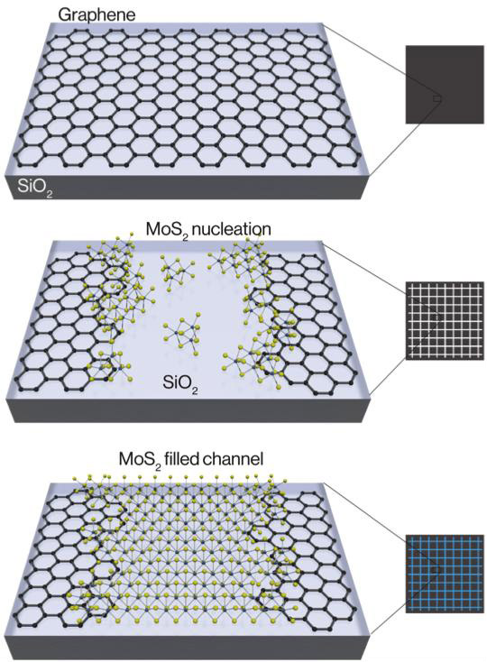
This schematic shows the chemical assembly of two-dimensional crystals. Graphene is first etched into channels and the TMDC molybdenum disulfide (MoS2) begins to nucleate around the edges and within the channel. On the edges, MoS2 slightly overlaps on top of the graphene. Finally, further growth results in MoS2 completely filling the channels. (credit: Berkeley Lab)

In an advance that helps pave the way for next-generation electronics and computing technologies — and possibly paper-thin devices — scientists with the U.S. Department of Energy’s Lawrence Berkeley National Laboratory (Berkeley Lab) have developed a way to chemically assemble transistors and circuits that are only a few atoms thick.

In addition, their method yields functional structures at a scale large enough to begin thinking about real-world applications and commercial scalability.

They reported their research online July 11 in the journal Nature Nanotechnology.

The scientists controlled the synthesis of a transistor in which narrow channels were etched onto conducting graphene, and a semiconducting material called a transition-metal dichalcogenide (TMDC) was seeded in the blank channels.

Both of these materials are single-layered crystals and atomically thin, so the two-part assembly yielded electronic structures that are essentially two-dimensional, and cover an area a few centimeters long and a few millimeters wide.

“This is a big step toward a scalable and repeatable way to build atomically thin electronics or pack more computing power in a smaller area,” says Xiang Zhang, a senior scientist in Berkeley Lab’s Materials Sciences Division who led the study.

Their work is part of a new wave of research aimed at keeping pace with Moore’s Law, which holds that the number of transistors in an integrated circuit doubles approximately every two years. To keep this pace, scientists predict that integrated electronics will soon require transistors that measure less than ten nanometers in length.

“With silicon, this will be extremely challenging, because the thickness of the transistor’s channel will become greater than the channel length, ultimately leading to difficult electrostatic control via the transistor gate,” the authors note. Nanomaterials such as inorganic nanowires and Stanford/IBM’s carbon nanotubes have been proposed, but require impractical “precise placement and orientation using complex fabrication techniques,” the authors point out.

The two-dimensional solution for keeping up with Moore’s law

Optical image of the atomically thin graphene–MoS heterostructure. Arrows indicate nucleation (junctions) with graphene of the MoS2 areas, forming transistors. (credit: Mervin Zhao et al./Nature Nanotechnology)

So researchers have now looked to two-dimensional crystals that are only one molecule thick as alternative materials to keep up with Moore’s Law. Using that approach, the Berkeley Lab scientists developed a way to seed a single-layered semiconductor — in this case, the TMDC molybdenum disulfide (MoS2) — into channels lithographically etched within a sheet of conducting graphene. The two atomic sheets meet to form nanometer-scale junctions that make atomically thin transistors in which the graphene conductor efficiently injects current into the MoS2.

“This approach allows for the chemical assembly of electronic circuits, using two-dimensional materials, which show improved performance compared to using traditional metals to inject current into TMDCs,” says Mervin Zhao, a lead author and Ph.D. student in Zhang’s group at Berkeley Lab and UC Berkeley.

The scientists demonstrated the usefulness of the structure by assembling it into the logic circuitry of an inverter (NOT gate). This further underscores the technology’s ability to lay the foundation for a chemically assembled atomic computer, the scientists say. They also note that the two-dimensional crystals were synthesized at a wafer scale, so the scalable design is compatible with current semiconductor manufacturing.

The research was supported by the Office of Naval Research and the National Science Foundation. Scientists from Cornell University were also involved in the research.


Abstract of Large-scale chemical assembly of atomically thin transistors and circuits

Next-generation electronics calls for new materials beyond silicon, aiming at increased functionality, performance and scaling in integrated circuits. In this respect, two-dimensional gapless graphene and semiconducting transition-metal dichalcogenides have emerged as promising candidates due to their atomic thickness and chemical stability. However, difficulties with precise spatial control during their assembly currently impede actual integration into devices. Here, we report on the large-scale, spatially controlled synthesis of heterostructures made of single-layer semiconducting molybdenum disulfide contacting conductive graphene. Transmission electron microscopy studies reveal that the single-layer molybdenum disulfide nucleates at the graphene edges. We demonstrate that such chemically assembled atomic transistors exhibit high transconductance (10 µS), on–off ratio (∼106) and mobility (∼17 cm2 V−1 s−1). The precise site selectivity from atomically thin conducting and semiconducting crystals enables us to exploit these heterostructures to assemble two-dimensional logic circuits, such as an NMOS inverter with high voltage gain (up to 70).

Allen Brain Observatory launched

The Allen Institute for Brain Science today announced the release of the Allen Brain Observatory.

This is standardized survey of cellular-level activity in the mouse visual system. The goal is to empower scientists to “investigate how circuits in the behaving mouse brain coordinate to drive activity and perception, and lays a crucial foundation for understanding perception, cognition and ultimately consciousness.”

The Allen Institute is known for its comprehensive brain atlases, with deep, high-quality data sets revealing where genes are expressed and how cells and connections are arranged in the mouse and human brain, according to Allan Jones, Ph.D., CEO of the Allen Institute.

With this new project, the researchers took a clever approach, combining a large variety of visual stimuli, including film clips and moving images, with the associated responses of neurons in four areas of the mouse visual cortex at multiple depths, sampling more than 18,000 neurons in total. The goal is to determine the “tuning,” or preference, of each individual neuron to visual features like motion and shape orientation, as well as complex images like natural scenes and movies that reveal integrative dynamics of visual processing.


Allen Institute | Allen Brain Observatory: Visualizing the brain in action

The data are presented as part of the suite of Allen Brain Atlas tools in the uniform and shareable Neurodata Without Borders file format, which allows scientists anywhere to easily mine and model the data. (The mouse is an important model system often used to understand the far less accessible and far larger human brain.) The data are presented in a novel visualization format through the Allen Brain Atlas data portal, and are accompanied by analysis tools and access to all raw data, which allows scientists to deeply explore the rules that govern how networks of cells in the visual cortex communicate.

“The Allen Brain Observatory is a stunning window into the visual brain of the mouse,” says Christof Koch, Ph.D., President and Chief Scientific Officer of the Allen Institute for Brain Science. “No one has ever taken this kind of standardized approach to surveying the active brain at cellular resolution in order to measure how the brain processes information in real time. This is a milestone in our quest to decode how the brain’s computations give rise to perception, behavior, and consciousness.

“Just like in astronomy, modelers and theoreticians worldwide can now study this wealth of data using their own analysis tools. If we want to understand higher-order brain functions, we need to understand not just the individual components of the brain but how they all work together.”

Understanding visual processing is a key gateway to understanding how other parts of the brain process information, and future releases of the Allen Brain Observatory will also explore the neural circuits that underlie more complex behaviors like decision-making, according to the researchers.


Allen Institute | Inside the Allen Brain Observatory