Surprising discovery of highly dynamic changes in olfactory region of the adult mouse brain

In light brown, in the center of the image, a new adult-born neuron. The neurons in blue are synaptic partner neurons, which connect to the new neurons. The neurons in dark brown are pre-existing neurons. (credit: Institut Pasteur/PM Lledo)

Scientists from the Institut Pasteur and the CNRS have made the surprising discovery that new neurons formed in the olfactory bulb of adult mice are constantly reorganizing the billions of synaptic contacts they establish among themselves (also described as constant structural plasticity).

The researchers found this puzzling because constant structural plasticity is normally confined to specific critical periods after birth, and “plasticity in neural circuits must strike a balance between flexibility and stability so new information can be acquired while previously learned information can be retained,” they note in a paper published in the journal Neuron.

The olfactory bulb in rodents and the hippocampus in humans are two of the areas of the brain capable of constantly regenerating their neurons in adulthood, but it had not been known that olfactory neurons were able to reorganize synaptic contacts.

Watching the brain change through a porthole

To observe the ongoing formation of neuronal circuits, the scientists marked the new granule cell (GC) inter-neurons with a green fluorescent protein (GFP), to view the dynamic changes, using two-photon microscopy imaging via a cranial window.

These experiments were carried out over a period of several months, following the entire life cycle of the new neurons. In the first three weeks of their life, these new neurons extended their cellular projections, known as dendrites, to form several ramifications, which subsequently became very stable.

Sample images of the same adult-born granule cell (GC) inter-neuron dendritic segment at a 2-day interval for 20–22, 40–42, and 60–62 days after virus injection (for monitoring) showing stable (closed arrowheads with numbers indexing stable spines), new (open arrowheads), and lost (asterisks) spines (credit: Kurt A. Sailor et al./Neuron)

They next observed neuronal spines — the structure where synapses form — and demonstrated that 20% of the synapses between new and pre-existing neurons were changed on a daily basis — a phenomenon that was also observed in their synaptic partners, the principal olfactory bulb neurons.

Adjusting efficiently and reliably to ongoing sensory changes

Using computer-based models, the authors showed that these dynamics enabled the synaptic network to adjust efficiently and reliably to ongoing sensory changes in the environment, enabling optimal processing of sensory information by the olfactory bulb.

“Our findings suggest that the plasticity of this constantly regenerating region of the brain occurs with continuous physical formation and elimination of synaptic connections. This structural plasticity reveals a unique dynamic mechanism that is vital for the regeneration and integration of new neurons within the adult brain circuit,” concluded the scientists.

More generally, they said, this study suggests a universal plasticity mechanism in brain regions that is closely associated with memory and learning.

This research was supported by the Institut Pasteur and the CNRS and was funded by AG2R-La Mondiale, the French National Research Agency, the “Revive” LabEx, and the “Biopsy” LabEx.


Abstract of Persistent Structural Plasticity Optimizes Sensory Information Processing in the Olfactory Bulb

In the mammalian brain, the anatomical structure of neural circuits changes little during adulthood. As a result, adult learning and memory are thought to result from specific changes in synaptic strength. A possible exception is the olfactory bulb (OB), where activity guides interneuron turnover throughout adulthood. These adult-born granule cell (GC) interneurons form new GABAergic synapses that have little synaptic strength plasticity. In the face of persistent neuronal and synaptic turnover, how does the OB balance flexibility, as is required for adapting to changing sensory environments, with perceptual stability? Here we show that high dendritic spine turnover is a universal feature of GCs, regardless of their developmental origin and age. We find matching dynamics among postsynaptic sites on the principal neurons receiving the new synaptic inputs. We further demonstrate in silico that this coordinated structural plasticity is consistent with stable, yet flexible, decorrelated sensory representations. Together, our study reveals that persistent, coordinated synaptic structural plasticity between interneurons and principal neurons is a major mode of functional plasticity in the OB.

How to bring the entire web to VR

Google is working on new features to bring the web to VR, according to Google Happiness Evangelist .

To help web developers embed VR content in their web pages, the Google Chromium team has been working towards WebVR support in Chromium (programmers: see Chromium Code Reviews), Beaufort said. That means you can now use Cardboard- or Daydream-ready VR viewers to see pages with compliant VR content while browsing the web with Chrome.

(credit: Google)

“The team is just getting started on making the web work well for VR so stay tuned, there’s more to come!” he said.

Google previously launched VR view,  which enables developers to embed immersive content on Android, iOS and the web. Users can view it on their phone, with a Cardboard viewer, or with a Chrome browser on their desktop computer.

For native apps, programmers can embed a VR view in an app or web page by grabbing the latest Cardboard SDK for Android or iOS and adding a few lines of code.

On the web, embedding a VR view is as simple as adding an iframe on your site, as KurzweilAI did in the 360-degrees view shown at the top of this page, using iframe code copied from the HTML on Introducing VR view: embed immersive content into your apps and websites on Google Developers Blog. (Chrome browser is required. In addition to a VR viewer, you can use either the mouse or the four arrow keys to explore the image in 360 degrees.)

A host of common chemicals endanger child brain development, NIH journal reports

(credit: Graphic by Julie McMahon)

In a new open-access report in the NIH journal Environmental Health Perspectives, 47 scientists, health practitioners, and children’s health advocates have made a consensus statement in “Project TENDR: Targeting Environmental Neuro-Developmental Risks“ — endorsed by nine medical organizations — and issued a call to action for renewed attention to the growing evidence that many common and widely available chemicals endanger neurodevelopment in fetuses and children of all ages.

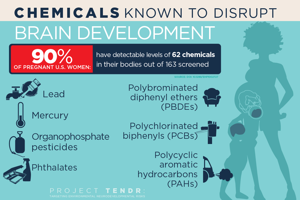
The list includes chemicals used extensively in consumer products and that have become widespread in the environment. Of most concern are lead and mercury; organophosphate pesticides used in agriculture and home gardens; phthalates, which are used in pharmaceuticals, plastics and personal care products; flame retardants known as polybrominated diphenyl ethers; and air pollutants produced by the combustion of wood and fossil fuels, said University of Illinois Comparative Biosciences professor Susan Schantz, one of dozens of individual signatories to the consensus statement.

The list provides “prime examples of toxic chemicals that can contribute to learning, behavioral, or intellectual impairment, as well as specific neurodevelopmental disorders such as ADHD or autism spectrum disorder,” according to the report.

Polychlorinated biphenyls

Polychlorinated biphenyls, once used as coolants and lubricants in transformers and other electrical equipment, also are of concern. PCBs were banned in the U.S. in 1977, but can persist in the environment for decades, she said.

“These chemicals are pervasive, not only in air and water, but in everyday consumer products that we use on our bodies and in our homes,” Schantz said. “Reducing exposures to toxic chemicals can be done, and is urgently needed to protect today’s and tomorrow’s children.”

“The human brain develops over a very long period of time, starting in gestation and continuing during childhood and even into early adulthood,” Schantz said. “But the biggest amount of growth occurs during prenatal development. The neurons are forming and migrating and maturing and differentiating. And if you disrupt this process, you’re likely to have permanent effects.”

Hormonal disrupters

Some of the chemicals of concern, such as phthalates and PBDEs, are known to interfere with normal hormone activity. For example, most pregnant women in the U.S. will test positive for exposure to phthalates and PBDEs, both of which disrupt thyroid hormone function.

“Thyroid hormone is involved in almost every aspect of brain development, from formation of the neurons to cell division, to the proper migration of cells and myelination of the axons after the cells are differentiated,” said Schantz. “It regulates many of the genes involved in nervous system development.”

Schantz and her colleagues at Illinois are studying infants and their mothers to determine whether prenatal exposure to phthalates and other endocrine disruptors leads to changes in the brain or behavior. This research, along with parallel studies in older children and animals, is a primary focus of the Children’s Environmental Health Research Center at Illinois, which Schantz directs.

Phthalates also interfere with steroid hormone activity. Studies link exposure to certain phthalates with attention deficits, lower IQ and conduct disorders in children. “Phthalates are everywhere; they’re in all kinds of different products. We’re exposed to them every day,” Schantz said.

The report criticizes current regulatory lapses that allow chemicals to be introduced into people’s lives with little or no review of their effects on fetal and child health. “For most chemicals, we have no idea what they’re doing to children’s neurodevelopment,” Schantz said. “They just haven’t been studied.

“And if it looks like something is a risk, we feel policymakers should be willing to make a decision that this or that chemical could be a bad actor and we need to stop its production or limit its use,” she said. “We shouldn’t have to wait 10 or 15 years — allowing countless children to be exposed to it in the meantime — until we’re positive it’s a bad actor.”

Project TENDR has a website with information about each of the chemicals of concern. The National Institute of Environmental Health Sciences at the National Institutes of Health and the U.S. Environmental Protection Agency fund the Children’s Environmental Health Research Center at the University of Illinois.

Project TENDR is an alliance of 48 of the nation’s top scientists, health professionals and health advocates. It was launched by Maureen Swanson of the Learning Disabilities Association of America and Irva Hertz-Picciotto of UC Davis, who brought together participants across many disciplines and sectors, including epidemiology, toxicology, exposure science, pediatrics, obstetrics and gynecology, nursing, public health, and federal and state chemical policy. Medical and scientific societies that have signed on in support include American Congress of Obstetricians and Gynecologists, American Nurses Association, Endocrine Society, National Association of Pediatric Nurse Practitioners, National Medical Association, National Hispanic Medical Association, Alliance of Nurses for Healthy Environments, Physicians for Social Responsibility and the National Council of Asian Pacific Island Physicians. TENDR’s long-term mission is to lower the incidence of neurodevelopmental disorders by reducing exposure levels to chemicals and pollutants that can contribute to these conditions, especially during fetal development and early childhood.


Steve Drake, Beckman Institute for Advanced Science and Technology | U. of I. biosciences professor Susan Schantz directs the Children’s Environmental Health Research Center at the University of Illinois, which is studying whether, and how, exposure to phthalates disrupts child brain development. Phthalates are used in some cosmetics, food packaging and products with fragrances.


Food Safety | From DDT to Glyphosate: Rachel Carson, We Need You Again


Abstract of Project TENDR: Targeting Environmental Neuro-Developmental Risks. The TENDR Consensus Statement

SUMMARY: Children in America today are at an unacceptably high risk of developing neurodevelopmental disorders that affect the brain and nervous system including autism, attention deficit hyperactivity disorder, intellectual disabilities, and other learning and behavioral disabilities. These are complex disorders with multiple causes—genetic, social, and environmental. The contribution of toxic chemicals to these disorders can be prevented. APPROACH: Leading scientific and medical experts, along with children’s health advocates, came together in 2015 under the auspices of Project TENDR: Targeting Environmental Neuro-Developmental Risks to issue a call to action to reduce widespread exposures to chemicals that interfere with fetal and children’s brain development. Based on the available scientific evidence, the TENDR authors have identified prime examples of toxic chemicals and pollutants that increase children’s risks for neurodevelopmental disorders. These include chemicals that are used extensively in consumer products and that have become widespread in the environment. Some are chemicals to which children and pregnant women are regularly exposed, and they are detected in the bodies of virtually all Americans in national surveys conducted by the U.S. Centers for Disease Control and Prevention. The vast majority of chemicals in industrial and consumer products undergo almost no testing for developmental neurotoxicity or other health effects. CONCLUSION: Based on these findings, we assert that the current system in the United States for evaluating scientific evidence and making health-based decisions about environmental chemicals is fundamentally broken. To help reduce the unacceptably high prevalence of neurodevelopmental disorders in our children, we must eliminate or significantly reduce exposures to chemicals that contribute to these conditions. We must adopt a new framework for assessing chemicals that have the potential to disrupt brain development and prevent the use of those that may pose a risk. This consensus statement lays the foundation for developing recommendations to monitor, assess, and reduce exposures to neurotoxic chemicals. These measures are urgently needed if we are to protect healthy brain development so that current and future generations can reach their fullest potential.

Prenatal exposure to acetaminophen may increase autism spectrum symptoms

Tylenol PM (left) and Tylenol (right) (credit: Ragesoss/CC)

A new study has found that paracetamol (aka acetaminophen; trade names include Tylenol and Panadol), which is used extensively during pregnancy, has a strong association with autism spectrum symptoms in boys and for both genders in relation to attention-related and hyperactivity symptoms.

The findings* were published this week in the International Journal of Epidemiology. This is the first study of its kind to report an independent association between the use of this drug in pregnancy and autism spectrum symptoms in children.

It is also the first study to report different effects on boys and girls. Comparing persistently to nonexposed children, the study has found an increase of 30 per cent in the risk of detriment to some attention functions, and an increase of two clinical symptoms of autism spectrum symptoms in boys. Boys also showed more autism spectrum symptoms when persistently exposed to paracetamol.

“Paracetamol could be harmful to neurodevelopment for several reasons,” said Co-author Dr. Jordi Júlvez, also a researcher at CREAL. “First of all, it relieves pain by acting on cannabinoid receptors in the brain.

Why boys are more likely to have autism

“Since these receptors normally help determine how neurons mature and connect with one another, paracetamol could alter these important processes. It can also affect the development of the immune system, or be directly toxic to some fetuses that may not have the same capacity as an adult to metabolize this drug, or by creating oxidative stress.”

There could also be an explanation for why boys are more likely to have autism spectrum symptoms: “The male brain may be more vulnerable to harmful influences during early life”, said Claudia Avella-Garcia. “Our differing gender results suggest that androgenic endocrine disruption, to which male brains could be more sensitive, may explain the association.”

The study concluded that the widespread exposure of infants to paracetamol in utero could increase the number of children with ADHD or autism spectrum symptoms. However, they stressed further studies should be conducted with more precise dosage measurements, and that the risks versus benefits of paracetamol use during pregnancy and early life should be assessed before treatment recommendations are made.

* Researchers in Spain recruited 2644 mother-child pairs in a birth cohort study during pregnancy. 88 per cent were evaluated when the child was one year old, and 79.9 per cent were evaluated when they were five years old. Mothers were asked about their use of paracetamol during pregnancy and the frequency of use was classified as never, sporadic, or persistent. Exact doses could not be noted due to mothers being unable to recall them exactly. 43 per cent of children evaluated at age one and 41 per cent assessed at age five were exposed to any paracetamol at some point during the first 32 weeks of pregnancy. When assessed at age five, exposed children were at higher risk of hyperactivity or impulsivity symptoms. Persistently exposed children in particular showed poorer performance on a computerised test measuring inattention, impulsivity and visual speed processing.


Abstract of Acetaminophen use in pregnancy and neurodevelopment: attention function and autism spectrum symptoms

Background: Acetaminophen is extensively used during pregnancy. But there is a lack of population-representative cohort studies evaluating its effects on a range of neuropsychological and behavioural endpoints. We aimed to assess whether prenatal exposure to acetaminophen is adversely associated with neurodevelopmental outcomes at 1 and 5 years of age.

Methods: This Spanish birth cohort study included 2644 mother-child pairs recruited during pregnancy. The proportion of liveborn participants evaluated at 1 and 5 years was 88.8% and 79.9%, respectively. Use of acetaminophen was evaluated prospectively in two structured interviews. Ever/never use and frequency of use (never, sporadic, persistent) were measured. Main neurodevelopment outcomes were assessed using Childhood Autism Spectrum Test (CAST), Conner’s Kiddie Continuous Performance Test (K-CPT) and ADHD-DSM-IV form list. Regression models were adjusted for social determinants and co-morbidities.

Results: Over 40% of mothers reported using acetaminophen. Ever-exposed offspring had higher risks of presenting more hyperactivity/impulsivity symptoms [incidence rate ratio (IRR) = 1.41, 95% confidence interval (CI) 1.01–1.98), K-CPT commission errors (IRR = 1.10, 1.03–1.17), and lower detectability scores (coefficient β = −0.75, −0.13–−0.02). CAST scores were increased in ever-exposed males (β = 0.63, 0.09–1.18). Increased effect sizes of risks by frequency of use were observed for hyperactivity/impulsivity symptoms (IRR = 2.01, 0.95–4.24) in all children, K-CPT commission errors (IRR = 1.32, 1.05–1.66) and detectability (β = −0.18, −0.36–0.00) in females, and CAST scores in males (β = 1.91, 0.44–3.38).

Conclusions: Prenatal acetaminophen exposure was associated with a greater number of autism spectrum symptoms in males and showed adverse effects on attention-related outcomes for both genders. These associations seem to be dependent on the frequency of exposure.

A spectacular survey of the distant universe

An image of a small section (0.4 percent) of the Ultra-Deep Survey field. Most of the objects in the image are very distant galaxies, observed as they were more than 9 billion years ago. In the full image, 250,000 galaxies have been detected over an area of sky four times the size of the full moon. (credit: Omar Almaini, University of Nottingham)

Astronomers at The University of Nottingham have released spectacular new infrared images of the distant Universe, providing the deepest view ever obtained over a large area of sky — showing more than 250,000 galaxies, including several hundred observed within the first billion years after the Big Bang.

The images are from the final data release from the Ultra-Deep Survey (UDS), which maps an area four times the size of the full Moon to unprecedented depth. Astronomers will use the new images to study the early stages of galaxy formation and evolution.

The release of the final UDS images represents the culmination of a project that began taking data in 2005. The scientists used the United Kingdom Infrared Telescope (UKIRT) on Hawaii to observe the same patch of sky repeatedly, building up more than 1000 hours of exposure time.

Understanding how galaxies evolved

Observing in the infrared is required for studying the distant Universe, since ordinary starlight is “redshifted” to longer wavelengths due to the cosmological expansion of the Universe.

Earlier releases of data from the UDS have already produced a wide range of scientific advances, including studies of the earliest galaxies in the first billion years after the Big Bang, measurements of the buildup of galaxies through cosmic time, and studies of the large-scale distribution of galaxies to weigh the mysterious “dark matter” that pervades the cosmos.

David Maltby, PhD, a postdoctoral research fellow at The University of Nottingham, said the scientists are aiming to understand how galaxies evolved to produce the rich diversity we see today.

“For example, we still don’t understand why the most massive galaxies are usually elliptical in shape, while less massive galaxies tend to be disk-shaped with spiral arms. By looking back in time to the early Universe we can catch these galaxies in their infancy, and observe them as they change and evolve over many billions of years.”

First self-driving vehicle death

Tesla Model S (credit: Tesla)

A Tesla Model S car was involved in a fatal crash yesterday, Tesla Motors announced today, June 30, on its blog.

“Joshua Brown, a 40-year-old Ohio owner of a Tesla Model S, died when his electric car drove under the trailer of an 18-wheel truck on a highway in Williston, Fla.,” The Wall Street Journal reports, based on local news reports.

“The vehicle was on a divided highway with Autopilot engaged when a tractor trailer drove across the highway perpendicular to the Model S,” according to the blog. “Neither Autopilot nor the driver noticed the white side of the tractor trailer against a brightly lit sky, so the brake was not applied. The high ride height of the trailer combined with its positioning across the road and the extremely rare circumstances of the impact caused the Model S to pass under the trailer, with the bottom of the trailer impacting the windshield of the Model S.”

The blog also noted that whenever autopilot is engaged, the car reminds the driver to “Always keep your hands on the wheel. Be prepared to take over at any time” and “makes frequent checks to ensure that the driver’s hands remain on the wheel and provides visual and audible alerts if hands-on is not detected. It then gradually slows down the car until hands-on is detected again.”

Tesla said it reported the accident to the National Highway Traffic Safety Administration immediately after learning about it yesterday.

Brown maintained a YouTube channel focused on the Tesla 7.0 Autopilot.

AI beats top U.S. Air Force tactical air combat experts in combat simulation

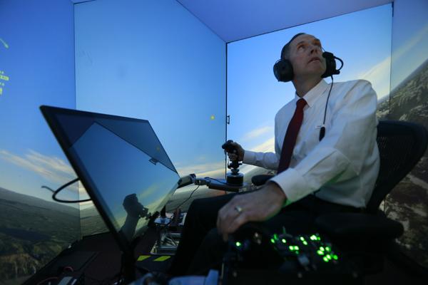
Retired U.S. Air Force Colonel Gene Lee, in a flight simulator, takes part in simulated air combat versus artificial intelligence technology developed by a team from industry, the U.S. Air Force, and University of Cincinnati. (credit: Lisa Ventre, University of Cincinnati Distribution A: Approved for public release; distribution unlimited. 88ABW Cleared 05/02/2016; 88ABW-2016-2270)

The U.S. Air Force got a wakeup call recently when AI software called ALPHA — running on a tiny $35 Raspberry Pi computer — repeatedly defeated retired U.S. Air Force Colonel Gene Lee, a top aerial combat instructor and Air Battle Manager, and other expert air-combat tacticians at the U.S. Air Force Research Lab (AFRL) in Dayton, Ohio. The contest was conducted in a high-fidelity air combat simulator.

According to Lee, who has considerable fighter-aircraft expertise (and has been flying in simulators against AI opponents since the early 1980s), ALPHA is “the most aggressive, responsive, dynamic and credible AI I’ve seen to date.” In fact, he was shot out of the air every time during protracted engagements in the simulator, he said.

ALPHA’s secret? Custom-designed “genetic fuzzy” algorithms designed for simulated air-combat missions, according to an open-access, unclassified paper published in the authoritative Journal of Defense Management. The paper was authored by a team of industry, Air Force, and University of Cincinnati researchers, including the AFRL Branch Chief.

ALPHA, which now runs on a standard consumer-grade PC, was developed by Psibernetix, Inc., an AFRL contractor founded by University of Cincinnati College of Engineering and Applied Science 2015 doctoral graduate Nick Ernest*, president and CEO of the firm, and a team of former Air Force aerial combat experts, including Lee.

“AI wingmen”

Today’s fighters close in on each other at speeds in excess of 1,500 miles per hour while flying at altitudes above 40,000 feet. The cost for a mistake is very high. Microseconds matter, but an average human visual reaction time is 0.15 to 0.30 seconds, and “an even longer time to think of optimal plans and coordinate them with friendly forces,” the researchers note in the paper.

Side view during active combat in simulator between two Blue (human-controlled) fighters vs. four Red (AI) fighters with >150 inputs but handicapped data sources. All Reds have successfully evaded missiles; one Blue has been destroyed. Blue AWACS [Airborne early warning and control system aircraft] shown in distance. (credit: Nicholas Ernest et al./Journal of Defense Management)

In fact, ALPHA works 250 times faster than humans, the researchers say. Nonetheless, ALPHA’s future role will stop short of fully autonomous combat.

According to the AFRL team, ALPHA will first be tested on “Unmanned Combat Aerial Vehicles (UCAV),” where ALPHA will be organizing data and creating a complete mapping of a combat scenario, such as a flight of four fighter aircraft — which it can do in less than a millisecond.

The AFRL team sees ACAVs as “AI wingmen” capable of engaging in air combat when teamed with manned aircraft.

The ACAVs will include an onboard battle management system able to process situational awareness, determine reactions, select tactics, and manage weapons — simultaneously evading dozens of hostile missiles, taking accurate shots at multiple targets, coordinating actions of squad mates, and recording and learning from observations of enemy tactics and capabilities.

Genetic fuzzy systems

The researchers based the design of ALPHA on a “genetic fuzzy tree” (GFT) — a subtype of “fuzzy logic” algorithms. The GFT is described in another open-access paper in Journal of Defense Management by Ernest and University of Cincinnati aerospace professor Kelly Cohen.

“Genetic fuzzy systems have been shown to have high performance, and a problem with four or five inputs can be solved handily,” said Cohen. “However, boost that to a hundred inputs, and no computing system on planet Earth could currently solve the processing challenge involved — unless that challenge and all those inputs are broken down into a cascade of sub-decisions.

“Most AI programming uses numeric-based control and provides very precise parameters for operations,” he said. In contrast, the AI algorithms that Ernest and his team developed are language-based, with if/then scenarios and rules able to encompass hundreds to thousands of variables. This language-based control, or fuzzy logic, can be verified and validated, Cohen says.

The “genetic” part of the “genetic fuzzy tree” system started with numerous automatically generated versions of ALPHA that proved themselves against a manually tuned version of ALPHA. The successful strings of code were then “bred” with each other, favoring the stronger, or highest performance versions.

In other words, only the best-performing code was used in subsequent generations. Eventually, one version of ALPHA rises to the top in terms of performance, and that’s the one that is utilized.

“In terms of emulating human reasoning, I feel this is to unmanned aerial vehicles as the IBM Deep Blue vs. Kasparov was to chess,” said Cohen. Or as Alpha Go was to Go.

* Support for Ernest’s doctoral research was provided  by the Dayton Area Graduate Studies Institute and the U.S. Air Force Research Laboratory.


UC | Flying in Simulator


Abstract of Genetic Fuzzy based Artificial Intelligence for Unmanned Combat Aerial Vehicle Control in Simulated Air Combat Missions

Breakthroughs in genetic fuzzy systems, most notably the development of the Genetic Fuzzy Tree methodology, have allowed fuzzy logic based Artificial Intelligences to be developed that can be applied to incredibly complex problems. The ability to have extreme performance and computational efficiency as well as to be robust to uncertainties and randomness, adaptable to changing scenarios, verified and validated to follow safety specifications and operating doctrines via formal methods, and easily designed and implemented are just some of the strengths that this type of control brings. Within this white paper, the authors introduce ALPHA, an Artificial Intelligence that controls flights of Unmanned Combat Aerial Vehicles in aerial combat missions within an extreme-fidelity simulation environment. To this day, this represents the most complex application of a fuzzy-logic based Artificial Intelligence to an Unmanned Combat Aerial Vehicle control problem. While development is on-going, the version of ALPHA presented within was assessed by Colonel (retired) Gene Lee who described ALPHA as “the most aggressive, responsive, dynamic and credible AI (he’s) seen-to-date.” The quality of these preliminary results in a problem that is not only complex and rife with uncertainties but also contains an intelligent and unrestricted hostile force has significant implications for this type of Artificial Intelligence. This work adds immensely to the body of evidence that this methodology is an ideal solution to a very wide array of problems.

A smarter ‘bionic’ cardiac patch that doubles as advanced pacemaker/arrhythmia detector

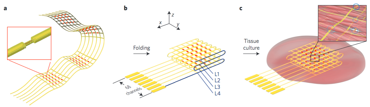
(a) Schematic of the free-standing macroporous nanoelectronic scaffold with nanowire FET (field effect transistor) arrays (red dots). Inset: one nanowire FET. (b) Folded 3D free-standing scaffolds with four layers of individually addressable FET sensors. (c) Schematic of nanoelectronic scaffold/cardiac tissue resulting from the culturing of cardiac cells within the 3D folded scaffold. Inset: the nanoelectronic sensors (blue circles) innervate the 3D cell network. (credit: Xiaochuan Dai at al./Nature Nanotechnology)

Harvard researchers have designed nanoscale electronic scaffolds (support structures) that can be seeded with cardiac cells to produce a new “bionic” cardiac patch (for replacing damaged cardiac tissue with pre-formed tissue patches). It also functions as a more sophisticated pacemaker: In addition to electrically stimulating the heart, the new design can change the pacemaker stimulation frequency and direction of signal propagation.

In addition, because because its electronic components are integrated throughout the tissue (instead of being located on the surface of the skin), it could detect arrhythmia far sooner, and “operate at far lower (safer) voltages than a normal pacemaker, [which] because it’s on the surface, has to use relatively high voltages,” according to Charles Lieber, the Mark Hyman, Jr. Professor of Chemistry and Chair of the Department of Chemistry and Chemical Biology.

Early arrhythmia detection, monitoring responses to cardiac drugs

“Even before a person started to go into large-scale arrhythmia that frequently causes irreversible damage or other heart problems, this could detect the early-stage instabilities and intervene sooner,” he said. “It can also continuously monitor the feedback from the tissue and actively respond.”

The patch might also find use, Lieber said, as a tool to monitor responses to cardiac drugs, or to help pharmaceutical companies screen the effectiveness of drugs under development.

In the long term, Lieber believes, the development of nanoscale tissue scaffolds represents a new paradigm for integrating biology with electronics in a virtually seamless way.

The bionic cardiac patch can also be a unique platform to study the tissue behavior evolving during some developmental processes, such as aging, ischemia, or differentiation of stem cells into mature cardiac cells.

Although the bionic cardiac patch has not yet been implanted in animals, “we are interested in identifying collaborators already investigating cardiac patch implantation to treat myocardial infarction in a rodent model,” he said. “I don’t think it would be difficult to build this into a simpler, easily implantable system.”

Could one day deliver cardiac patch/pacemaker via injection

Using the injectable electronics technology he pioneered last year, Lieber even suggested that similar cardiac patches might one day simply be delivered by injection. “It may actually be that, in the future, this won’t be done with a surgical patch,” he said. “We could simply do a co-injection of cells with the mesh, and it assembles itself inside the body, so it’s less invasive.”

“I think one of the biggest impacts would ultimately be in the area that involves replacement of damaged cardiac tissue with pre-formed tissue patches,” Lieber said. “Rather than simply implanting an engineered patch built on a passive scaffold, our work suggests it will be possible to surgically implant an innervated patch that would now be able to monitor and subtly adjust its performance.”

In the long term, Lieber believes, the development of nanoscale tissue scaffolds represents a new paradigm for integrating biology with electronics in a virtually seamless way.

The study is described in a June 27 paper published in Nature Nanotechnology.


Abstract of Three-dimensional mapping and regulation of action potential propagation in nanoelectronics-innervated tissues

Real-time mapping and manipulation of electrophysiology in three-dimensional (3D) tissues could have important impacts on fundamental scientific and clinical studies, yet realization is hampered by a lack of effective methods. Here we introduce tissue-scaffold-mimicking 3D nanoelectronic arrays consisting of 64 addressable devices with subcellular dimensions and a submillisecond temporal resolution. Real-time extracellular action potential (AP) recordings reveal quantitative maps of AP propagation in 3D cardiac tissues, enable in situtracing of the evolving topology of 3D conducting pathways in developing cardiac tissues and probe the dynamics of AP conduction characteristics in a transient arrhythmia disease model and subsequent tissue self-adaptation. We further demonstrate simultaneous multisite stimulation and mapping to actively manipulate the frequency and direction of AP propagation. These results establish new methodologies for 3D spatiotemporal tissue recording and control, and demonstrate the potential to impact regenerative medicine, pharmacology and electronic therapeutics.

This remarkable fish has no cortex but intelligently switches between electrical and vision senses

The elephantnose fish (Gnathonemus petersii) explores objects in its surroundings by using its eyes or its electrical sense (credit: Timo Moritz)

With its tiny brain (and no cortex), the elephantnose fish (Gnathonemus petersii)* achieves performance comparable to that of humans or other mammals in certain tasks, according to zoologists at the University of Bonn and a colleague from Oxford.

To perceive objects in the water, the fish uses electrolocation (similar to the echolocation of bats) to perceive objects in the water, aided by an electrical organ in its tail, which emits electrical impulses, and numerous electrical sensor organs in its skin. It also uses its visual sense.

Curiously, in an experiment** in which the animals became familiar with an object in an aquarium with the visual sense, they were also able to recognize it again using the electrical sense, although they had never perceived it electrically before.

Experimental setup. Aquarium tanks were divided into two compartments by a partition with two closable gates, behind which were positioned two objects (indicated in red) 1 cm from the gate. Distance grids, which were placed directly behind the gates and which could be passed by pushing them aside, made sure that the fish kept the correct minimal distance to the objects. The larger compartment (experimental area) was again divided into two compartments, one gate leading to each compartment. (credit: S. Schumacher et al./PNAS)

In the experiment, when the two senses delivered different information in the close range of up to two centimeters, the fish trusted only the electrical information and were then “blind” to the visual stimuli. In contrast, for more distant objects, the animals relied above all on their eyes. And they perceived the environment best by using their visual and electrical senses in combination.

“This ability has only been found in mammals, suggesting such a high-level function might be associated with complex mammalian brain structures. Furthermore, the modality-specific inputs are weighted dynamically according to their reliability at different ranges,” the researchers note in a paper published online on PNAS.

“A transfer between the different senses was previously known only for certain highly developed mammals, such as monkeys, dolphins, rats, and humans”, says Professor Gerhard von der Emde at the Institute of Zoology at the University of Bonn. “In a dark, unfamiliar apartment, people feel their way forward to avoid stumbling. When the light goes on, the obstacles felt are recognized by the eye without any problem.”

The secret is in the cerebellum

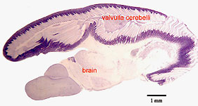
Parasagittal cresyl violet stained section through the brain of a Gnathonemus petersii showing the topography of the valvulla cerebelli covering most of the rest of the brain.
“Brain” on this figure includes, from left to right, parts of the telencephalon, diencephalon and mesencephalon. (credit: E. Gilissen, based on the material of Prof. J.M. Allman, Caltech)

So exactly how does Gnathonemus petersii achieve this surprising level of intelligence? A clue is provided by Emmanuel Gilissen on the website of The Royal Museum for Central Africa. He explains that the African mormyrids or elephant-nose fishes were noted for having unusually large brains already more than a century ago. “For a mean body mass of 26 g, the mean brain weight of Gnathonemus petersii reaches 0.53 g, almost three times its expected mean value of 0.19 g, as calculated from the relationship between brain size and body size in teleost fish (Kaufman, 2003).

“This character is probably, at least in part, related to their ability to sense prey and to communicate by generating and perceiving electric fields (Nieuwenhuys and Nicholson, 1969).  In contrast with mammals, it is the cerebellum, and not the telencephalon that is greatly enlarged in these fishes. … In elephant-nose fishes, the valvula cerebelli covers most of the rest of the brain.  In contrast, in another highly derived brain such as the human brain, it is the telencephalon, and more specifically the neocortex, a telencephalic structure unique to mammals, that entirely covers the rest of the brain. …

“In the electric fish Gnathonemus petersii, the brain is responsible for approximately 60% of body O2 consumption, a figure three times higher than that for any other vertebrate studied so far, including human.”

So are these electrically genius fish up there with the macaw?

* Gnathonemus petersii is widespread in the flowing waters of West Africa and hunts insect larva at dawn and dusk.

** The elephantnose fish was in an aquarium connected to two different chambers; the animal could choose. Behind openings to the chambers there were differently shaped objects: a sphere or a cuboid. The fish learned to steer toward one of these objects by being rewarded with insect larvae. Subsequently, it searched for this object again, to obtain the reward again. When does the fish use a particular sense? To answer this question, the researchers repeated the experiments in absolute darkness. Now the fish could rely only on its electrical sense. As shown by images taken with an infrared camera, it was able to recognize the object only at short distances. With the light on the fish was most successful, because it was able to use its eyes and the electrical sense for the different distances. To find out when the fish used its eyes alone, the researchers made the objects invisible to the electrical sense. Now, the sphere and cuboid to be discriminated had the same electrical characteristics as the water.


Abstract of Cross-modal object recognition and dynamic weighting of sensory inputs in a fish

Most animals use multiple sensory modalities to obtain information about objects in their environment. There is a clear adaptive advantage to being able to recognize objects cross-modally and spontaneously (without prior training with the sense being tested) as this increases the flexibility of a multisensory system, allowing an animal to perceive its world more accurately and react to environmental changes more rapidly. So far, spontaneous cross-modal object recognition has only been shown in a few mammalian species, raising the question as to whether such a high-level function may be associated with complex mammalian brain structures, and therefore absent in animals lacking a cerebral cortex. Here we use an object-discrimination paradigm based on operant conditioning to show, for the first time to our knowledge, that a nonmammalian vertebrate, the weakly electric fish Gnathonemus petersii, is capable of performing spontaneous cross-modal object recognition and that the sensory inputs are weighted dynamically during this task. We found that fish trained to discriminate between two objects with either vision or the active electric sense, were subsequently able to accomplish the task using only the untrained sense. Furthermore we show that cross-modal object recognition is influenced by a dynamic weighting of the sensory inputs. The fish weight object-related sensory inputs according to their reliability, to minimize uncertainty and to enable an optimal integration of the senses. Our results show that spontaneous cross-modal object recognition and dynamic weighting of sensory inputs are present in a nonmammalian vertebrate.

Artificial synapse said to rival biological synapses in energy consumption and function

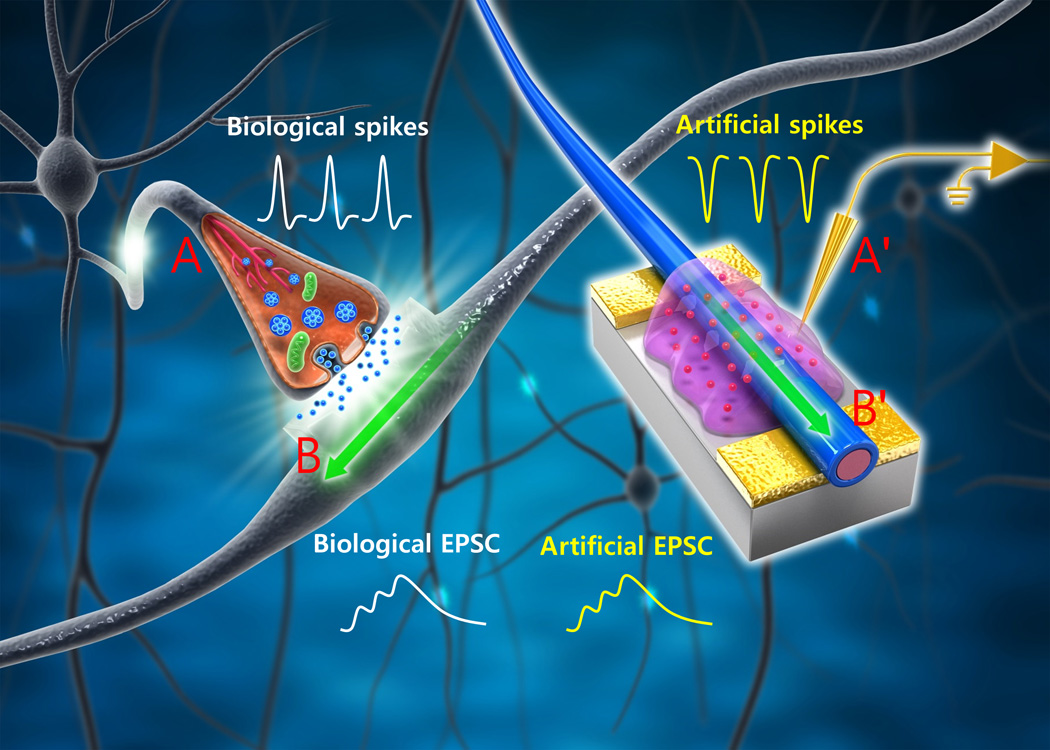
Schematic of biological neuronal network and an organic nanowire (ONW) synaptic transistor (ST) that emulates a biological synapse. The yellow conductive lines and probe (A′) mimic an axon (A) that delivers presynaptic spikes from a pre-neuron to the presynaptic membrane. The mobile ions in the ion gel move in the electrical field, analogous to the biological neuron transmitters in the synaptic cleft; the field later induces an excitatory postsynaptic current (EPSC, light blue line) in the biological dendrite (B). An ONW (B′) combined with a drain electrode (yellow surface) mimics a biological dendrite (B). EPSC (light green line) is generated in the ONW in response to presynaptic spikes and is delivered to a post-neuron through connections to the drain electrode. (credit: Wentao Xu et al./Science Advances)

An artificial synapse that emulates a biological synapse while requiring less energy has been developed by Pohang University Of Science & Technology (POSTECH) researchers* in Korea.

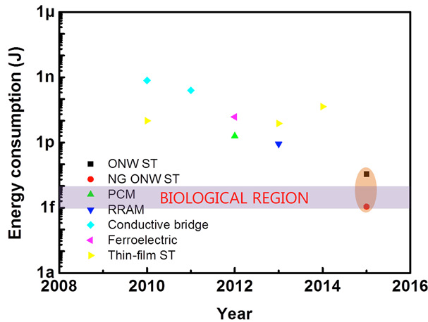
Energy consumption in Joules per synaptic event of currently available synaptic devices.*** NG, nanogap; PCM, phase change memory; RRAM, resistive switching random access memory. (credit: Wentao Xu et al./Science Advances)

A human synapse consumes an extremely small amount of energy (~10 fJ or femtojoules** per synaptic event).

The researchers have fabricated an organic nanofiber (ONF), or organic nanowire (ONW), electronic device that emulates the important working principles and energy consumption of biological synapses while requiring only ~1 fJ per synaptic event. The ONW also emulates the morphology (form) of a synapse.

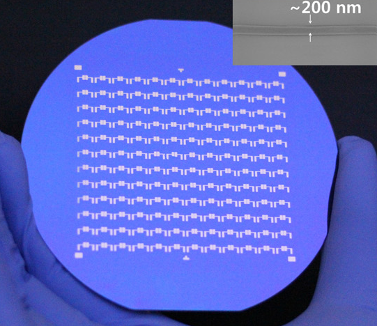
Array of 144 ONW STs (organic nanowire synaptic transistors) fabricated on a 4-inch silicon wafer. Inset: Scanning electron microscopy (SEM) image of a typical ONW with a diameter of 200 nm. (credit: Wentao Xu et al./Science Advances)

The morphology of ONFs is similar to that of nerve fibers, which form crisscrossing grids to enable the high memory density of a human brain. The researchers say the highly-aligned ONFs can be massively produced with precise control over alignment and dimension; and this morphology may make possible the future construction of the high-density memory of a neuromorphic (brain-form-emulating) system.****

The researchers say they have emulated important working principles of a biological synapse, such as paired-pulse facilitation (PPF), short-term plasticity (STP), long-term plasticity (LTP), spike-timing dependent plasticity (STDP), and spike-rate dependent plasticity (SRDP).

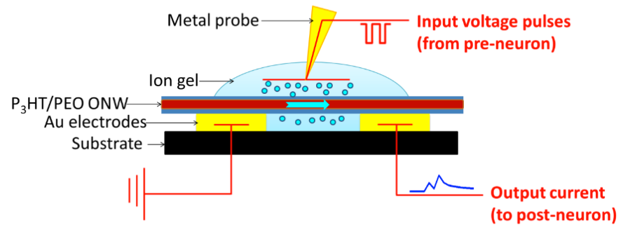
The ONW STs are three-terminal devices. Input voltage pulses that emulate presynaptic spikes from a pre-neuron are applied to the metal probe, which functions as a gate electrode. The input pulses cause ions to migrate in the ion gel, causing a change in the source-drain (yellow rectangles) current flowing through the semiconducting ONW (red). The output current signal — which emulates post-synaptic current flowing to a post-neuron in a biological synapse — is measured (during characterization) by recording the drain current. (credit: Wentao Xu et al./Science Advances)

The artificial synapse devices provide a new research direction in neuromorphic electronics and open a new era of organic electronics with high memory density and low energy consumption, the researchers claim. Potential applications include neuromorphic computing systems, AI systems for self-driving cars, analysis of big data, cognitive systems, robot control, medical diagnosis, stock-trading analysis, remote sensing, and other smart human-interactive systems and machines in the future, they suggest.

This research was supported by the Pioneer Research Center Program and Center for Advanced Soft-Electronics as a Global Frontier Project, funded by the Korean Ministry of Science, ICT, and Future Planning.

The research was published an open-access paper in Science Advances, a new sister journal of Science.

* Prof. Tae-Woo Lee, Wentao Xu, and Sung-Yong Min, PhD, with the Dept. of Materials Science and Engineering at POSTECH

** A fJ (femtojoule) is 10-15 Joule (one watt-second). 

*** ~1014 synapses.

**** Previous attempts to realize synaptic functions in single electronic devices include resistive random access memory (RRAM), phase change memory (PCM), conductive bridges, and synaptic transistors.


Abstract of Organic core-sheath nanowire artificial synapses with femtojoule energy consumption

Emulation of biological synapses is an important step toward construction of large-scale brain-inspired electronics. Despite remarkable progress in emulating synaptic functions, current synaptic devices still consume energy that is orders of magnitude greater than do biological synapses (~10 fJ per synaptic event). Reduction of energy consumption of artificial synapses remains a difficult challenge. We report organic nanowire (ONW) synaptic transistors (STs) that emulate the important working principles of a biological synapse. The ONWs emulate the morphology of nerve fibers. With a core-sheath–structured ONW active channel and a well-confined 300-nm channel length obtained using ONW lithography, ~1.23 fJ per synaptic event for individual ONW was attained, which rivals that of biological synapses. The ONW STs provide a significant step toward realizing low-energy–consuming artificial intelligent electronics and open new approaches to assembling soft neuromorphic systems with nanometer feature size.