How birds unlock their ultraviolet vision super-sense

Some birds have been found to be as intelligent as mammals. And some that can see ultraviolet (UV) light live in a super-sensory world apart, able to transmit and receive signals between each other in a way that is invisible to many other species.

Now the ability of finches, sparrows, and many other birds to see ultraviolet (UV) light is explained in a study published in the journal eLife by scientists at the Washington University School of Medicine in St. Louis.

Carotenoid pigments determine bird’s UV perception or not

The study reveals two essential adaptions that enable birds to expand their vision into the UV range: chemical changes in light-filtering pigments called carotenoids (such as carotene, found in carrots, associated with vitamin A) and the tuning of light-sensitive proteins called opsins (light-sensitive proteins), some of which are also used in optogenetics research.

Birds acquire carotenoids through their diets and process them in a variety of ways to shift their light absorption toward longer (visible light) or shorter (UV) wavelengths.

“There are two types of light-sensitive cells, called photoreceptors, in the eye: rods and cones. Cone photoreceptors are responsible for color vision. While humans have blue, green, and red-sensitive cones only, birds have a fourth cone type which is either violet or UV-sensitive, depending on the species,” says senior author Joseph Corbo, MD, PhD, Associate Professor of Pathology and Immunology.

UV vs visual perception by cones in the eye is fine-tuned by evolution

“Our approach showed that blue-cone sensitivity is fine-tuned through a change in the chemical structure of carotenoid pigments within the photoreceptor, allowing both violet and UV-sighted birds to maximize how many colors they can see.”

The study also revealed that sensitivity of the violet/UV cone and the blue cone in birds must move in sync to allow for optimum vision. Among bird species, there is a strong relationship between the light sensitivity of opsins within the violet/UV cone and mechanisms within the blue cone, which coordinate to ensure even UV vision.

Taken together, these results suggest that both blue and violet cone cells have adapted during evolution to enhance color vision in birds.

Birds have achieved UV vision by use of a specialized optical organelle, the pigmented cone oil droplet. These oil droplets are located in the path of light through the receptor and act as cutoff filters matched to the visual pigment sensitivity of each cone subtype.

Spectral filtering in bird cones. a) A flat-mounted chicken retina under brightfield illumination that shows the distinctive pigmentation of the cone oil droplets. (b) A diagram of the avian single cone photoreceptors showing the relative position of the oil droplet within the cells (top) and a representation of the spectral filtering cutoff effects of the droplet (bottom). (credit: Matthew B Toomey et al./eLife)

“The majority of bird species rely on vision as their primary sense, and color discrimination plays a crucial role in their essential behaviors, such as choosing mates and foraging for food. This explains why birds have evolved one of the most richly endowed color vision systems among vertebrates,” says first author Matthew Toomey, a postdoctoral fellow at the Washington University School of Medicine.

“The precise coordination of sensitivity and filtering in the visual system may, for example, help female birds discriminate very fine differences in the elaborate coloration of their suitors and choose the fittest mates. This refinement of visual sensitivity could also facilitate the search for hidden seeds, fruits, and other food items in the environment.”

The team now plans to investigate the underlying molecular mechanisms that help modify the carotenoid pigments and light-sensitive protein tuning in a wide range of bird species, to gather further insights into the evolution of UV vision.


Abstract of Complementary shifts in photoreceptor spectral tuning unlock the full adaptive potential of ultraviolet vision in birds

Color vision in birds is mediated by four types of cone photoreceptors whose maximal sensitivities (λmax) are evenly spaced across the light spectrum. In the course of avian evolution, the λmax of the most shortwave-sensitive cone, SWS1, has switched between violet (λmax > 400 nm) and ultraviolet (λmax < 380 nm) multiple times. This shift of the SWS1 opsin is accompanied by a corresponding short-wavelength shift in the spectrally adjacent SWS2 cone. Here, we show that SWS2 cone spectral tuning is mediated by modulating the ratio of two apocarotenoids, galloxanthin and 11’,12’-dihydrogalloxanthin, which act as intracellular spectral filters in this cell type. We propose an enzymatic pathway that mediates the differential production of these apocarotenoids in the avian retina, and we use color vision modeling to demonstrate how correlated evolution of spectral tuning is necessary to achieve even sampling of the light spectrum and thereby maintain near-optimal color discrimination.

FDA places hold on clinical trial of cancer treatment previously reported on KurzweilAI — UPDATE: FDA releases hold

Juno Therapeutics, Inc. announced July 7 that it has received notice from the FDA that it has placed a clinical hold on an immune-cell cancer treatment known as the “ROCKET” trial, which was reported on KurzweilAI on Mar. 10, 2016.

The clinical hold was initiated after two patient deaths, which followed the recent addition of fludarabine to the pre-conditioning regimen. Juno has proposed to the FDA to continue the ROCKET trial using JCAR015 with cyclophosphamide pre-conditioning alone.

Further information on Forbes.

UPDATE JULY 13, 2016: Juno Therapeutics announced Tuesday July 12 that the FDA has removed the hold and “the ROCKET trial will continue enrollment using JCAR015 with cyclophosphamide pre-conditioning only.” In related news, the Law Offices of Howard G. Smith announced an investigation on behalf of investors of Juno Therapeutics, Inc. (NASDAQ: JUNO) concerning the Company and its officers’ possible violations of federal securities laws and noted that “Juno’s share price fell significantly in value, dropping almost 32%.”

How to detect early signs of Alzheimer’s with a simple eye exam before symptoms appear

Monochromatic images of normal mouse retina at four wavelengths from blue (upper left) to red (lower right). Boxes in lower right show locations typical of amyloid (a substance found in the brain associated with Alzheimer’s). (credit: Swati S. More et al./IOVS)

University of Minnesota (UMN) scientists and associates have developed new technology that can detect signs of Alzheimer’s before the onset of symptoms — early enough to give drugs a chance to work — in mice and humans by simply examining the back of their eyes.

Looking at Alzheimer’s effects through the eye is a key advantage of the new technology. “The retina of the eye is not just ‘connected’ to the brain — it is part of the central nervous system,” said Swati More, PhD, of the Center for Drug Design at UMN, co-author of a paper recently published in Investigative Ophthalmology & Visual Science (IOVS).

The brain and retina undergo similar changes due to Alzheimer’s disease, he said, but “unlike the brain, the retina is easily accessible to us. We saw changes in the retinas of Alzheimer’s mice before the typical age at which neurological signs are observed.”

Human clinical trials are set to start in July to test the technology in humans who are 40–75 years old (for more information on participating in the clinical trial, you can visit the trial website).

Optical spectra recorded from human and mouse retina samples. Upper two lines show the intensity of light for different wavelengths of light from red (right side) to blue (left side) for a normal human retina (heavy line) and for an Alzheimer’s (AD) patients retina. Comparable mouse plots, shown below (WT = normal mouse; APP1/PS1 = Alzheimer’s model mouse), show a similar pattern. (credit: Swati S. More et al./IOVS)

Early detection of Alzheimer’s is critical for two reasons. “First, effective treatments need to be administered well before patients show actual neurological signs,” said co-author Robert Vince, PhD, of the Center for Drug Design at the University of Minnesota (UMN). “Second, since there are no available early detection techniques, drugs currently cannot be tested to determine if they are effective against early Alzheimer’s disease. An early diagnostic tool like ours could help the development of drugs as well.”


Abstract of Early Detection of Amyloidopathy in Alzheimer’s Mice by Hyperspectral Endoscopy

Purpose: To describe a spectral imaging system for small animal studies based on noninvasive endoscopy of the retina, and to present time-resolved spectral changes from live Alzheimer’s mice prior to cognitive decline, corroborating our previous in vitro findings.

Methods: Topical endoscope fundus imaging was modified to use a machine vision camera and tunable wavelength system for acquiring monochromatic images across the visible to near-infrared spectral range. Alzheimer’s APP/PS1 mice and age-matched, wild-type mice were imaged monthly from months 3 through 8 to assess changes in the fundus reflection spectrum. Optical changes were fit to Rayleigh light scatter models as measures of amyloid aggregation.

Results: Good quality spectral images of the central retina were obtained. Short-wavelength reflectance from Alzheimer’s mice retinae showed significant reduction over time compared to wild-type mice. Optical changes were consistent with an increase in Rayleigh light scattering in neural retina due to soluble Aβ1–42aggregates. The changes in light scatter showed a monotonic increase in soluble amyloid aggregates over a 6-month period, with significant build up occurring at 7 months.

Conclusions: Hyperspectral imaging technique can be brought inexpensively to the study of retinal changes caused by Alzheimer’s disease progression in live small animals. A similar previous finding of reduction in the light reflection over a range of wavelengths in isolated Alzheimer’s mice retinae, was reproducible in the living Alzheimer’s mice. The technique presented here has a potential for development as an early Alzheimer’s retinal diagnostic test in humans, which will support the treatment outcome.

Locusts engineered as biorobotic sensing machines

Sensors placed on the insect monitor neural activity while they are freely moving, decoding the odorants present in their environment. (credit: Baranidharan Raman)

Washington University in St. Louis engineers have developed an innovatiave “bio-hybrid nose” that could be used in homeland security applications, such as detecting explosives, replacing state-of-the-art miniaturized chemical sensing devices limited to a handful of sensors.

Compare that to the locust antenna (where their chemical sensors are located): “it has several hundreds of thousands of sensors and of a variety of types,” says Baranidharan Raman, associate professor of biomedical engineering, who has received a three-year, $750,000 grant from the Office of Naval Research (ONR).

The team previously found that locusts can correctly identify a particular odor, even with other odors present — and even in complex situations, such as overlapping with other scents or in different background conditions.

Replacing canines

In previous research, the opening of the locust maxillary palps to the trained odorant was used as an indicator of acquired memory. The palps were painted with non-odorous organic-chemical green paint to facilitate tracking. (credit: Debajit Saha et al./Nature Communications)

The ingenious idea in the new study by the Raman Lab is to remotely monitor neural activity from the insect brain while they are freely moving, exploring, and decoding the odorants present in their environment, which will require innovative low-power electronic components to collect, log, and transmit data.

The locusts could also collect samples using remote control. To do that, the engineers are developing a plasmonic “tattoo” made of a biocompatible silk to apply to the locusts’ wings. It will generate mild heat to help steer locusts to move toward particular locations by remote control. The tattoos, studded with plasmonic nanostructures, also can collect samples of volatile organic compounds in their proximity, which would allow the researchers to conduct secondary analysis of the chemical makeup of the compounds using more conventional methods.

“The canine olfactory system still remains the state-of-the-art sensing system for many engineering applications, including homeland security and medical diagnosis,” Raman said. “However, the difficulty and the time necessary to train and condition these animals, combined with lack of robust decoding procedures to extract the relevant chemical sending information from the biological systems, pose a significant challenge for wider application.

This deadly soil bug can reach your brain in a day, end up in spinal cord

B. pseudomallei soil-dwelling bacterium endemic in tropical and subtropical regions worldwide, particularly in Thailand and northern Australia (credit: Wikipedia CC)

Imagine a  deadly bacteria that can be picked up by a simple sniff and can travel to your brain and spinal cord in just 24 hours. Or one that could just be quietly sitting there, waiting for an opportune moment. Or maybe just doing small incremental damage ever day over a lifetime … as you lose the function in your brain incrementally.

That’s the grisly finding (in mice), published in Immunity and Infection this week, of a new study by Australian Griffith University and Bond University scientists.

The pathogenic bacteria Burkholderia pseudomallei, which causes the potentially fatal disease melioidosis, kills 89,000 people around the world each year and is prevalent in northern Australia, where a person with melioidosis has a 20–50 per cent chance of dying once it infects the brain. The bacterium is found in the northern parts of the Northern Territory, including Darwin.

In Southeast Asia 50 per cent of the population may be positive for melioidosis, and in places like Cambodia the mortality rate is as high as 50 per cent.

But for the rest of us, the findings could also lead to discoveries of how the common staphylococcus and acne bacterium also end up in the spinal cord, as well as how chlamydia travels to the brain in Alzheimer’s patients. Or even explain common back problems, which could be where bacteria have infected your bone, causing pain that could be simply treated with antibiotics, according to the researchers.

Tracing the bacteria in mice brains and spinal cords

(Top) A schematic drawing of a mouse brain showing the location of various images. (Bottom) D: A B. pseudomallei rod (arrow) present in trigeminal nerve near the connection between the trigeminal nerve in the brain and the brainstem. (E) B. pseudomallei rod (arrow) with a fluorescent particle (arrow with tail) after the merge between the trigeminal nerve and brainstem. Scale bars in μm. (credit: James A. St John et al./Infection and Immunity)

The olfactory mucosa, located in the nose, is very close to the brain and it has long been known that viruses could reach the brain from the olfactory mucosa. But researchers have not understand exactly how the bacteria traveled to the brain and spinal cord, or just how quickly.

To find out, James St. John, PhD., Head of Griffith’s Clem Jones Centre for Neurobiology and Stem Cell Research, and associates infected mice with B. pseudomallei. They were able to trace the bacteria travels from the nerves in the nasal cavity before moving to the brain stem and then into the spinal cord. (He noted that this could also be a pathway for many other common bacteria.)

“Our latest results represent the first direct demonstration of transit of a bacterium from the olfactory mucosa to the central nervous system (CNS) via the trigeminal nerve; bacteria were found a considerable distance from the olfactory mucosa, in the brain stem, and even more remarkably in the spinal cord,” said professor Ifor Beacham from the Griffith Institute for Glycomics.

Researchers will now work on ways to stimulate supporting cells that could remove the bacteria. St. John said the work was important because the bacteria had the potential to be used as a bioweapon and knowing how to combat it was extremely important.

“Bacteria have been implicated as a major causative agent of some types of back pain. We now need to work out whether the bacteria that cause back pain also can enter the brainstem and spinal cord via the trigeminal nerve,” he added.


Abstract of Burkholderia pseudomallei rapidly infects the brainstem and spinal cord via the trigeminal nerve after intranasal inoculation

Infection with Burkholderia pseudomallei causes melioidosis, a disease with a high mortality rate (20% in Australia and 40% in south-east Asia). Neurological melioidosis is particularly prevalent in northern Australian patients and involves brainstem infection, which can progress to the spinal cord; however, the route by which the bacteria invade the central nervous system (CNS) is unknown. We have previously demonstrated that B. pseudomallei can infect the olfactory and trigeminal nerves within the nasal cavity following intranasal inoculation. As the trigeminal nerve projects into the brainstem, we investigated whether the bacteria could continue along this nerve to penetrate the CNS. After intranasal inoculation of mice, B. pseudomallei caused low-level localised infection within the nasal cavity epithelium, prior to invasion of the trigeminal nerve in small numbers. B. pseudomallei rapidly invaded the trigeminal nerve and crossed the astrocytic barrier to enter the brainstem within 24 hours and then rapidly progressed over 2000 μm into the spinal cord. To rule out that the bacteria used a haematogenous route, we used a capsule-deficient mutant of B. pseudomallei, which does not survive in the blood, and found that it also entered the CNS via the trigeminal nerve. This suggests that the primary route of entry is via the nerves that innervate the nasal cavity. We found that actin-mediated motility could facilitate initial infection of the olfactory epithelium. Thus, we have demonstrated that B. pseudomallei can rapidly infect the brain and spinal cord via the trigeminal nerve branches that innervate the nasal cavity.

Neurons grown from stem cells in a dish reveal clues about autism

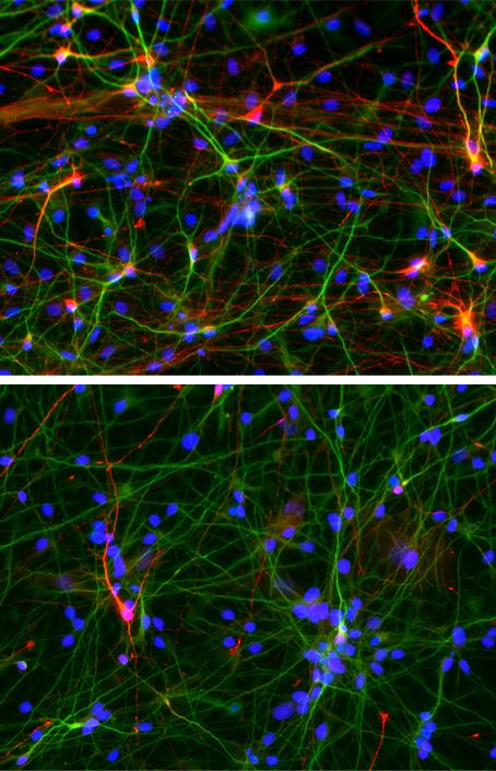
Salk researchers have turned the skin cells of people with autism spectrum disorder into neurons. These cells show specific defects (indicated by red and green dots in the neuron) compared with neurons derived from healthy people, including diminished ability to form excitatory connections with other neurons  (credit: Salk Institute)

Why do the brains of up to 30 percent of people with autism spectrum disorder grow faster than usual, early in life? A new study co-led by Salk Institute scientists has used a new stem cell reprogramming technique to find out.

Published July 6, 2016 in the journal Molecular Psychiatry, the Salk team found that stem cell-derived neurons made fewer connections in a dish compared to cells from healthy individuals. The scientists were also able to restore communication between the cells by adding IGF-1, a drug currently being evaluated in clinical trials of autism.*

Neurons derived from people with autism spectrum disorder, shown in the bottom panel, form fewer inhibitory connections, shown in the red stain, compared to those derived from healthy individuals (top panel). The total number of neurons that researchers were able to generate was about the same between the two groups. (credit: Salk Institute)

Autism affects approximately 1 out of every 68 children in the United States. It is characterized by problems communicating, difficulties interacting with others, and in repetitive behaviors, although the symptoms range dramatically in type and severity. There is no known cause of autism, according to the scientists.

Led by senior investigator Rusty Gage, a professor in Salk’s Laboratory of Genetics and holder of the Vi and John Adler Chair for Research on Age-Related Neurodegenerative Diseases, the researchers created stem cells from a subset of people with autism whose brains had grown as much as 23 percent faster than usual, but had subsequently normalized.

Improvement by adding IGF-1

The neuron precursor cells derived from the patients multiplied faster than those of typically developing individuals. The finding supports a theory some experts have put forth that brain enlargement is caused by disruptions to the cell’s normal cycle of division, the researchers say. In addition, the stem cell-derived neurons of individuals with autism behaved abnormally, bursting with activity less often compared with those cells of healthy people.

Those neurons’ activity seemed to improve by adding IGF-1, which is known to enhance the connections between neurons. The group plans to use the patient cells to investigate the molecular mechanisms behind IGF-1’s effects, in particular probing for changes in gene expression with treatment.

Other authors on the study include researchers from the University of California, San Francisco; the University of California, Los Angeles; the University of California San Diego; Case Western Reserve University, University of Rochester School of Medicine and Dentistry, and Icahn School of Medicine at Mount Sinai.

The research was supported by the California Institute for Regenerative Medicine, the National Institutes of Health, The International Rett Syndrome Foundation, a NARSAD Independent Investigator Award, a NIMH Autism Center of Excellence Program Project grant, The Leona M. and Harry B. Helmsley Charitable Trust, The JPB Foundation, the Robert and Mary Jane Engman Foundation, the CDMRP Autism Research Program, the University of California, San Diego Clinical and Translational Research Institute, and Autism Speaks.

* In 2010, Gage, Carol Marchetto of Salk’s Laboratory of Genetics, Alysson Muotri of the University of California, San Diego, and their collaborators showed they could recreate features of Rett syndrome—a rare disorder that shares features of autism but is caused by mutations in a single gene—in a petri dish.

They did so by taking skin cells from patients, adding a mix of chemicals that instructed those cells to form stem cells, and in turn, coaxing their new stem cells into neurons. The ability to form what’s called induced pluripotent stem cells (iPSCs) from human cells was pioneered by researchers in 2007, but some scientists were initially skeptical that the new technology could lend insight into complex heritable disorders such as autism.

“In that study, induced pluripotent stem cells gave us a window into the birth of a neuron that we would not otherwise have,” says Marchetto, a senior staff scientist and the study’s first author. “Seeing features of Rett syndrome in a dish gave us the confidence to next study classical autism.”


Abstract of Altered proliferation and networks in neural cells derived from idiopathic autistic individuals

Autism spectrum disorders (ASD) are common, complex and heterogeneous neurodevelopmental disorders. Cellular and molecular mechanisms responsible for ASD pathogenesis have been proposed based on genetic studies, brain pathology and imaging, but a major impediment to testing ASD hypotheses is the lack of human cell models. Here, we reprogrammed fibroblasts to generate induced pluripotent stem cells, neural progenitor cells (NPCs) and neurons from ASD individuals with early brain overgrowth and non-ASD controls with normal brain size. ASD-derived NPCs display increased cell proliferation because of dysregulation of a β-catenin/BRN2 transcriptional cascade. ASD-derived neurons display abnormal neurogenesis and reduced synaptogenesis leading to functional defects in neuronal networks. Interestingly, defects in neuronal networks could be rescued by insulin growth factor 1 (IGF-1), a drug that is currently in clinical trials for ASD. This work demonstrates that selection of ASD subjects based on endophenotypes unraveled biologically relevant pathway disruption and revealed a potential cellular mechanism for the therapeutic effect of IGF-1.

Molecular flip in crystals driven by light creates microrobotic propulsion

(a) Sequential micrographs of one cycle of self-oscillation observed under 435-nm light and schematic illustrations. (b) Schematic illustration showing setup. (credit: Tomonori Ikegami et al./Angewandte Chemie)

Hokkaido University researchers have designed a crystal material that continually flips between two positions like a paddle, propelling an attached structure, when stimulated by blue light. It could lead to bio-inspired microrobots that deliver drugs to target tissues, for example.

The team made azobenzene-oleic acid crystals, composed of an organic compound called azobenzene, commonly used in dye manufacturing, and oleic acid, commonly found in cooking oil. Azobenzene molecules take two structurally different forms: cis and trans, and they were found to switch back and forth when stimulated by the light.

The frequency of the motion also increased with increased light intensity. Some crystal complexes they created even exhibited swimming-like motions in the water, the researchers report. Previously reported light-responsive materials have been limited in their ability to deform, the researchers noted.

“The importance of this study lies in the realization of macroscopic self-oscillation by the repeated reversible reaction of a molecular machine with the cooperative transformation of a molecular assembly,” the researchers note in a paper published in the journal Angewandte Chemie. “These results provide a fundamental strategy for constructing dynamic self-organizations in supramolecular systems to achieve bioinspired molecular systems.”


Ikegami T. et. al. | A crystalline assembly of azobenzene derivative and oleate showed oscillatory bending-unbending motion under continuous 435-nm light irradiation.


Abstract of Dissipative and Autonomous Square-Wave Self-Oscillation of a Macroscopic Hybrid Self-Assembly under Continuous Light Irradiation

Building a bottom-up supramolecular system to perform continuously autonomous motions will pave the way for the next generation of biomimetic mechanical systems. In biological systems, hierarchical molecular synchronization underlies the generation of spatio-temporal patterns with dissipative structures. However, it remains difficult to build such self-organized working objects via artificial techniques. Herein, we show the first example of a square-wave limit-cycle self-oscillatory motion of a noncovalent assembly of oleic acid and an azobenzene derivative. The assembly steadily flips under continuous blue-light irradiation. Mechanical self-oscillation is established by successively alternating photoisomerization processes and multi-stable phase transitions. These results offer a fundamental strategy for creating a supramolecular motor that works progressively under the operation of molecule-based machines.

Facebook’s Secret Conversations

(credit: Facebook)

Facebook began today (Friday, July 8) rolling out a new beta-version feature for Messenger called “Secret Conversations,” allowing for “one-to-one secret conversations … that will be end-to-end encrypted and which can only be read on one device of the person you’re communicating with.”

Facebook suggests the feature will be useful for discussing an illness or sending financial information (as in the pictures above).  You can choose to set a timer to control the length of time each message you send remains visible within the conversation. (Rich content like GIFs, videos, and making payments are not supported.)

The technology, described in a technical whitepaper (open access), is based on the Signal Protocol developed by Open Whisper Systems, which is also used in Open Whisper Systems’ own Signal messaging app (Chrome, iOS, Android),  WhatsApp, and Google’s Allo (not yet launched).

Unlike WhatsApp and iMessage, which automatically encrypt every message, Secret Conversations only works from a single device and is opt-in, which “will likely rankle many privacy advocates,” says Wired .

But not as much as all of these encrypted services rankle law enforcement agencies, since the feature hampers surveillance capabilities, it adds.


Robot mimics vertebrate motion

Pleurobot (credit: EPFL)

École polytechnique fédérale de Lausanne (EPFL) scientists have invented a new robot called “Pleurobot” that mimics the way salamanders walk and swim with unprecedented detail.

Aside from being cool (and a likely future Disney attraction), the researchers believe designing the robot will provide a new tool for understanding the evolution of vertebrate locomotion. That could lead to better understanding of how the spinal cord controls the body’s locomotion, which may help develop future therapies and neuroprosthetic devices for paraplegic patients and amputees.

Pleurobot mimics Salamander. Neurobiologists say electrical stimulation of the spinal cord is what determines whether the salamander walks, crawls or swims: At lowest level of stimulation, the salamander walks; with higher stimulation, its pace increases, and beyond some threshold the salamander begins to swim. (credit: EPFL)

Simulating the 3D motion of the salamander’s locomotion requires exceptional precision. The Biorobotics Laboratory scientists started by shooting detailed x-ray videos of the salamander species Pleurodeles waltl from the top and the side, tracking up to 64 points along its skeleton while it performed different types of motion in water and on the ground.

Auke Ijspeert and his team at EPFL then 3D-printed bones and motorized joints, and even created a “nervous system” using electronic circuitry, allowing the Pleurobot to walk, crawl, and even swim underwater.*

Ijspeert thinks that the design methodology used for the Pleurobot can help develop other types of “biorobots,” which could become important tools in neuroscience and biomechanics.

The research, described in the Royal Society journal Interface, received funding from the Swiss National Center of Competence in Research (NCCR) in Robotics and from the Swiss National Science Foundation.


École polytechnique fédérale de Lausanne | A new robot mimics vertebrate motion

* In the design process, the researchers identified the minimum number of motorized segments required, as well as the optimal placement along the robot’s body, to replicate many of the salamander’s types of movement. That made it possible to construct Pleurobot with fewer bones and joints than the real-life creature — only 27 motors and 11 segments along its spine (the real animal has 40 vertebrae and multiple joints, some of which can even rotate freely and move side-to-side or up and down). 


Abstract of From cineradiography to biorobots: an approach for designing robots to emulate and study animal locomotion

Robots are increasingly used as scientific tools to investigate animal locomotion. However, designing a robot that properly emulates the kinematic and dynamic properties of an animal is difficult because of the complexity of musculoskeletal systems and the limitations of current robotics technology. Here, we propose a design process that combines high-speed cineradiography, optimization, dynamic scaling, three-dimensional printing, high-end servomotors and a tailored dry-suit to construct Pleurobot: a salamander-like robot that closely mimics its biological counterpart, Pleurodeles waltl. Our previous robots helped us test and confirm hypotheses on the interaction between the locomotor neuronal networks of the limbs and the spine to generate basic swimming and walking gaits. With Pleurobot, we demonstrate a design process that will enable studies of richer motor skills in salamanders. In particular, we are interested in how these richer motor skills can be obtained by extending our spinal cord models with the addition of more descending pathways and more detailed limb central pattern generator networks. Pleurobot is a dynamically scaled amphibious salamander robot with a large number of actuated degrees of freedom (DOFs: 27 in total). Because of our design process, the robot can capture most of the animal’s DOFs and range of motion, especially at the limbs. We demonstrate the robot’s abilities by imposing raw kinematic data, extracted from X-ray videos, to the robot’s joints for basic locomotor behaviours in water and on land. The robot closely matches the behaviour of the animal in terms of relative forward speeds and lateral displacements. Ground reaction forces during walking also resemble those of the animal. Based on our results, we anticipate that future studies on richer motor skills in salamanders will highly benefit from Pleurobot’s design.

New tech could have helped police locate shooters in Dallas

Potential shooter location in Dallas (credit: Fox News)

JULY 8, 3:56 AM EDT — Livestreamed data from multiple users with cell phones and other devices could be used to help police locate shooters in a situation like the one going on right now in Dallas, says Jon Fisher, CEO of San Francisco-based CrowdOptic.

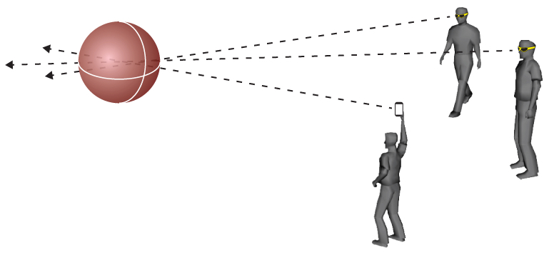
Here’s how it would work: You view (or record a video of) a shooter with your phone. Your location and the direction you are facing is now immediately available on your device and could be coordinated with data from other persons at the scene to triangulate the position of the shooter.

A CrowdOptic “cluster” with multiple people focused on the same object (credit: CrowdOptic)

This technology, called the “CrowdOptic Interactive Streaming platform,” is already in place, using Google Glass livestreaming, in several organizations, including UCSF Medical Center, Denver Broncos, and Futton, Inc. (working with Chinese traffic police).

Fisher told KurzweilAI his company’s software is also integrated with Cisco Jabber livestreaming video and conferencing products (and soon Spark), and with Sony SmartEyeglass, and that iOS and Android apps are planned.

CrowdOptic also has a product called CrowdOptic Eye, a “powerful, low-bandwidth live streaming device designed to … broadcast live video and two-way audio from virtually anywhere.”

“We’re talking about phones now, but think about all other devices, such as drones, that will be delivering these feeds to CNN and possibly local police,” he said.

ADDED July 11:

“When all attempts to negotiate with the suspect, Micah Johnson, failed under the exchange of gunfire, the Department utilized the mechanical tactical robot, as a last resort, to deliver an explosion device to save the lives of officers and citizens. The robot used was the Remotec, Model  F-5, claw and arm extension with an explosive device of C4 plus ‘Det’ cord.  Approximate weight of total charge was one pound.” — Statement July 9, 2016 by Dallas police chief David O. Brown

The Dallas police department’s decision to use a robot to kill the shooter Thursday July 7, raises questions. For example: Why wasn’t a non-lethal method used with the robot, such as a tranquilizer dart, which also might have given police an opportunity to acquire more information, including the location of claimed bombs and cohorts possibly associated with the crime?