Gene circuits in live cells that perform complex analog/digital computations

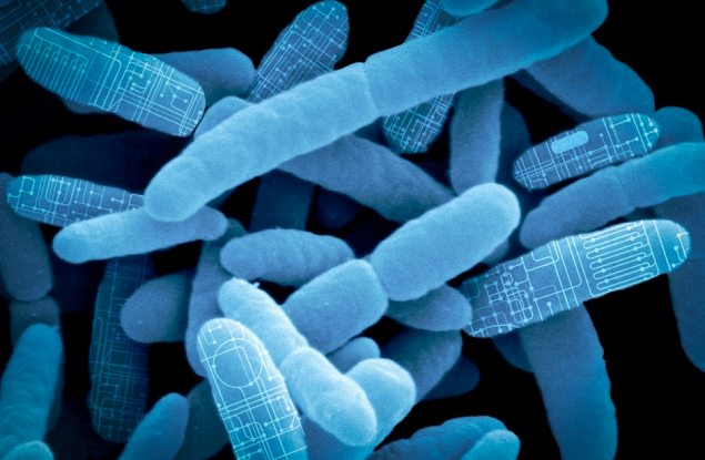
MIT researchers have developed synthetic biological circuits (from bacteria, for example, as shown here) that combine analog and digital computation as “living therapeutics” to treat major diseases and rare genetic disorders (credit: Synlogic)

MIT researchers have developed synthetic biological circuits that combine both analog (continuous) and digital (discrete) computation — allowing living cells to carry out complex processing operations, such as releasing a drug in response to low glucose levels.

The research is presented in an open-access paper published in the journal Nature Communications.


Background: analog vs. digital biological circuits

Like electronic circuits, living cells are capable of performing computations that are either continuous (analog) — like the way eyes adjust to gradual changes in the light levels — or digital, involving simple discrete on or off processes, such as a cell’s self-programmed death (apoptosis). Current synthetic biological systems, in contrast, have tended to focus on either analog or digital processing, limiting the range of uses.
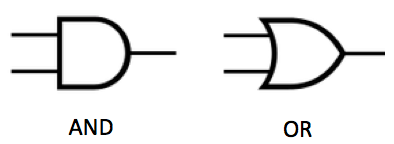
Two basic logic circuits. An AND gate fires only if both inputs are “true” (for example, both inputs have a 1 volt signal, not zero). An OR gate fires if either (or both) of the inputs is true (for example, the top input has a 1 volt signal and the bottom input has zero. (credit: KurzweilAI)

Digital systems are based on a simple binary output, such as 0 or 1, so performing complex computational operations requires the use of a large number of parts (such as AND and OR logic gates) to make the decision, which is difficult to achieve in synthetic biological systems. (There are seven basic logic gates: AND, OR, XOR, NOT, NAND, NOR, and XNOR, as explained here.)

Using genes (instead of voltages), synthetic biologists design genetic circuits (arrangements of DNA components) that can perform new functions. For example, here’s a gene circuit that was constructed using Escherichia coli bacteria (source: Imperial College London/Nature Communications study):
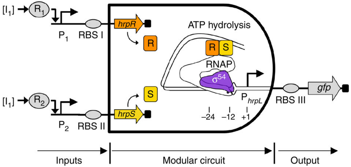
Example of a biological AND gate. Two environment-responsive gene promoters (a region of DNA that initiates transcription — that is, copying a particular segment of DNA into RNA), P1 and P2, act as the inputs to drive the transcriptions of hrpR and hrpS genes, and respond to small molecules. Transcription of the output promoter gfp is turned on only when both proteins HrpR and HrpS are present. (credit: Baojun Wang et al./Nature Communications)


“Most of the work in synthetic biology has focused on the digital approach, because [digital systems] are much easier to program,” says Timothy Lu, an associate professor of electrical engineering and computer science and of biological engineering, and head of the Synthetic Biology Group at MIT’s Research Laboratory of Electronics.

The new synthetic circuits can measure the level of an analog input, such as a particular chemical relevant to a disease, and then make a binary decision — for example, turning on an output, such as a drug that treats the disease if the level is in the right range.

The new circuits are based on multiple elements. For example, a threshold module consists of a sensor that detects analog levels of a particular chemical, which controls the expression of the second digital component, a recombinase gene, which can then switch on or off a segment of DNA by converting it into a digital (on or off) output. (This conversion process is similar to electronic devices known as comparators, which take analog input signals and convert them into a digital output.)

If the concentration of the chemical reaches a certain level, the threshold module expresses the recombinase gene, causing it to flip the DNA segment (which contains a gene or gene-regulatory element, which then alters the expression of a desired output).

“So this is how we take an analog input, such as a concentration of a chemical, and convert it into a 0 or 1 signal,” Lu says. “And once that is done, and you have a piece of DNA that can be flipped upside down, then you can put together any of those pieces of DNA to perform digital computing,” he says.

Ternary logic for three-way glucose decisions

The team has also built an analog-to-digital converter circuit that implements ternary (three-valued) logic. The circuit, which is capable of producing two different outputs, will only switch on in response to either a high or low concentration range of an input.

In the future, the circuit could be used to detect glucose levels in the blood and respond in one of three ways depending on the concentration, he says. “If the glucose level was too high, you might want your cells to produce insulin, if the glucose was too low you might want them to make glucagon, and if it was in the middle you wouldn’t want them to do anything,” he says.

Similar analog-to-digital converter circuits could also be used to detect a variety of chemicals, simply by changing the sensor, Lu says.

Detecting inflammation and environmental conditions

The researchers are investigating the idea of using analog-to-digital converters to detect levels of inflammation in the gut caused by inflammatory bowel disease, for example, and releasing different amounts of a drug in response.

Immune cells used in cancer treatment could also be engineered to detect different environmental inputs, such as oxygen or tumor lysis (cell breakdown) levels, and vary the immune-call therapeutic activity in response.

Other research groups are also interested in using the devices for environmental applications, such as engineering cells that detect concentrations of water pollutants, Lu says.

The research team recently created a spinout company, called Synlogic, which is now attempting to use simple versions of the circuits to engineer probiotic bacteria that can treat diseases in the gut. The company hopes to begin clinical trials of these bacteria-based treatments within the next 12 months.


Abstract of Synthetic mixed-signal computation in living cells

Living cells implement complex computations on the continuous environmental signals that they encounter. These computations involve both analogue- and digital-like processing of signals to give rise to complex developmental programs, context-dependent behaviours and homeostatic activities. In contrast to natural biological systems, synthetic biological systems have largely focused on either digital or analogue computation separately. Here we integrate analogue and digital computation to implement complex hybrid synthetic genetic programs in living cells. We present a framework for building comparator gene circuits to digitize analogue inputs based on different thresholds. We then demonstrate that comparators can be predictably composed together to build band-pass filters, ternary logic systems and multi-level analogue-to-digital converters. In addition, we interface these analogue-to-digital circuits with other digital gene circuits to enable concentration-dependent logic. We expect that this hybrid computational paradigm will enable new industrial, diagnostic and therapeutic applications with engineered cells.

Are you smarter than a macaw?

The macaw has a brain the size of an unshelled walnut, compared to the macaque monkey’s lemon-sized brain. But the macaw has more neurons in its forebrain — the portion of the brain associated with intelligent behavior — than the macaque. (credit: Vanderbilt University)

The first study to systematically measure the number of neurons in the brains of more than two dozen species of birds has found that the birds that were studied consistently have more neurons packed into their small brains than those in mammalian or even primate brains of the same mass.

The study results were published online in an open-access paper in the Proceedings of the National Academy of Sciences early edition on the week of June 13.

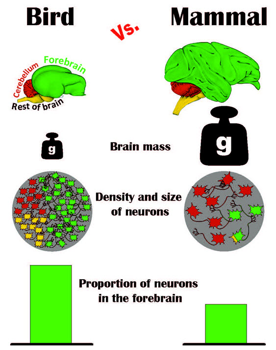
Graphic summary of the results of the avian brain study (credit: Pavel Nemec, Charles University of Prague)

“For a long time having a ‘bird brain’ was considered to be a bad thing. Now it turns out that it should be a compliment,” said Vanderbilt University neuroscientist Suzana Herculano-Houzel, senior author of the paper with Pavel Němec at the Charles University in Prague.

The study answers a puzzle that comparative neuroanatomists have been wrestling with for more than a decade: How can birds with their small brains perform complicated cognitive behaviors?

The conundrum was created by a series of studies beginning in the previous decade that directly compared the cognitive abilities of parrots and crows with those of primates. The studies found that the birds could manufacture and use tools, use insight to solve problems, make inferences about cause-effect relationships, recognize themselves in a mirror, and plan for future needs, among other cognitive skills previously considered the exclusive domain of primates.

The collection of avian brains that the scientists analyzed. For each species, the total number of neurons (in millions) in each brain is shown in yellow, the number of neurons (in millions) in the forebrain (pallium) is shown in blue and the brain mass (in grams) is shown in red. The scale bar in the lower right is 10 mm. (credit: Suzana Herculano-Houzel, Vanderbilt University)

So scientists assumed avian brains must  be wired differently from primate brains. Two years ago, even this hypothesis was knocked down by a detailed study of pigeon brains, which concluded that they are, in fact, organized along quite similar lines to those of primates.

More neurons in the forebrain than previously thought

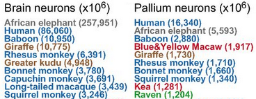
Top ten in number of whole-brain neurons and pallium (forebrain) neurons for the avian and mammalian species examined (credit: Seweryn Olkowicz et al./PNAS)

The new study provides a plausible explanation: Birds can perform these complex behaviors because birds’ forebrains contain a lot more neurons than any one had previously thought — as many as in mid-sized primates.

“We found that birds, especially songbirds and parrots, have surprisingly large numbers of neurons in their pallium: the part of the brain that corresponds to the cerebral cortex, which supports higher cognition functions such as planning for the future or finding patterns. That explains why they exhibit levels of cognition at least as complex as primates,” said Herculano-Houzel.

That’s because the neurons in avian brains are much smaller and more densely packed than those in mammalian brains, the study found. Parrot and songbird brains, for example, contain about twice as many neurons as primate brains of the same mass and two to four times as many neurons as equivalent rodent brains.

Also, the proportion of neurons in the forebrain is significantly higher, the study found.

More than one way to build better brains

“In designing brains, nature has two parameters it can play with: the size and number of neurons and the distribution of neurons across different brain centers,” said Herculano-Houzel, “and in birds we find that nature has used both of them.”

Although she acknowledges that the relationship between intelligence and neuron count has not yet been firmly established, Herculano-Houzel and her colleagues argue that having the same or greater forebrain neuron counts than primates with much larger brains can potentially provide the birds with much higher “cognitive power” per pound than mammals.

In other words, there’s more than one way to build better brains. Previously, neuroanatomists thought that as brains grew larger, neurons had to grow bigger as well because they had to connect over longer distances. “But bird brains show that there are other ways to add neurons: Keep most neurons small and locally connected and only allow a small percentage to grow large enough to make the longer connections. This keeps the average size of the neurons down,” she explained.

But that raises troubling questions:

  • Does the surprisingly large number of neurons in bird brains comes at a correspondingly large energetic cost?
  • Are the small neurons in bird brains a response to selection for small body size due to flight, or possibly the ancestral way of adding neurons to the brain — from which mammals, not birds, may have diverged.

Herculano-Houzel hopes that the results of the study and the questions it raises will stimulate other neuroscientists to begin exploring the mysteries of the avian brain, especially how their behavior compares to that of mammals of similar numbers of neurons or brain size.

Researchers at Charles University in Prague and the University of Vienna were also involved in the study.


Vanderbilt University | Bird Brain: Smarter Than You Think


Vanderbilt University | Study gives new meaning to the term “bird brain”


Abstract of Birds have primate-like numbers of neurons in the forebrain

Some birds achieve primate-like levels of cognition, even though their brains tend to be much smaller in absolute size. This poses a fundamental problem in comparative and computational neuroscience, because small brains are expected to have a lower information-processing capacity. Using the isotropic fractionator to determine numbers of neurons in specific brain regions, here we show that the brains of parrots and songbirds contain on average twice as many neurons as primate brains of the same mass, indicating that avian brains have higher neuron packing densities than mammalian brains. Additionally, corvids and parrots have much higher proportions of brain neurons located in the pallial telencephalon compared with primates or other mammals and birds. Thus, large-brained parrots and corvids have forebrain neuron counts equal to or greater than primates with much larger brains. We suggest that the large numbers of neurons concentrated in high densities in the telencephalon substantially contribute to the neural basis of avian intelligence.

Mobilizing mitochondria to regenerate damaged neurons

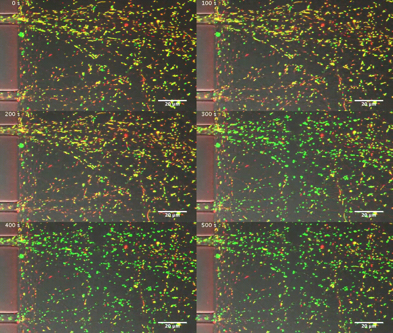
After axonal injury, nearby mitochondria become incapable of producing ATP, as indicated by their change in color from yellow (healthy) to green (damaged) (credit: Zhou et al., 2016)

Boosting the transport of mitochondria (cell energy suppliers) along neuronal axons enhances the ability of mouse nerve cells to repair themselves and regrow after injury or disease, researchers at the National Institute of Neurological Disorders and Stroke report in The Journal of Cell Biology.

Neurons need large amounts of energy to extend their axons long distances through the body. This energy — in the form of adenosine triphosphate (ATP) — is provided by mitochondria.

During development, mitochondria are transported up and down growing axons to generate ATP wherever it is needed. In adults, however, mitochondria become less mobile as mature neurons produce a protein called syntaphilin that anchors the mitochondria in place.

Zu-Hang Sheng and colleagues at the National Institute of Neurological Disorders and Stroke wondered whether this decrease in mitochondrial transport might explain why adult neurons are typically unable to regrow after injury.

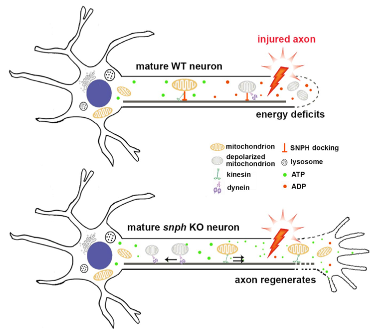
Repairing damaged neurons (top) by enhancing mitochondrial transport (bottom) (credit: Bing Zhou et al./JCB)

They initially found that when mature mouse axons are severed, nearby mitochondria are damaged and become unable to provide sufficient ATP to support injured nerve regeneration. However, when the researchers experimentally removed syntaphilin from the nerve cells (by using a genetically modified mouse), mitochondrial transport was enhanced, allowing the damaged mitochondria to be replaced by healthy mitochondria capable of producing ATP.

The Syntaphilin-deficient mature neurons therefore regained the ability to regrow after injury, just like young neurons.

“Our in vivo and in vitro studies suggest that activating an intrinsic growth program requires the coordinated modulation of mitochondrial transport and recovery of energy deficits. Such combined approaches may represent a valid therapeutic strategy to facilitate regeneration in the central and peripheral nervous systems after injury or disease,” Sheng says.


Abstract of Facilitation of axon regeneration by enhancing mitochondrial transport and rescuing energy deficits

Although neuronal regeneration is a highly energy-demanding process, axonal mitochondrial transport progressively declines with maturation. Mature neurons typically fail to regenerate after injury, thus raising a fundamental question as to whether mitochondrial transport is necessary to meet enhanced metabolic requirements during regeneration. Here, we reveal that reduced mitochondrial motility and energy deficits in injured axons are intrinsic mechanisms controlling regrowth in mature neurons. Axotomy induces acute mitochondrial depolarization and ATP depletion in injured axons. Thus, mature neuron-associated increases in mitochondria-anchoring protein syntaphilin (SNPH) and decreases in mitochondrial transport cause local energy deficits. Strikingly, enhancing mitochondrial transport via genetic manipulation facilitates regenerative capacity by replenishing healthy mitochondria in injured axons, thereby rescuing energy deficits. An in vivo sciatic nerve crush study further shows that enhanced mitochondrial transport in snph knockout mice accelerates axon regeneration. Understanding deficits in mitochondrial trafficking and energy supply in injured axons of mature neurons benefits development of new strategies to stimulate axon regeneration.

Higher intake of whole grains associated with lower risk of major chronic diseases and death

Cereal plant, 7-grain bread, wholegrain food (credit: iStock)

A meta-analysis of 45 studies (64 publications) of consumption of whole grain by an international team of researchers, led by Dagfinn Aune, PhD, at Imperial College London, found lower risks of coronary heart disease and cardiovascular disease overall, as well as deaths from all causes and from specific diseases, including stroke, cancer, diabetes, infectious and respiratory diseases.

The researchers say these results “strongly support dietary recommendations to increase intake of whole grain foods in the general population to reduce risk of chronic diseases and premature mortality.”

The results have been published in an open-access paper in the British Medical Journal (BMJ).

The greatest benefit was seen for people who increased from no intake of whole grain to two servings per day, equivalent to 32 g/day, such as 32 g of whole grain wheat, or to 60 g product/day, such as 60 g of whole grain wheat bread.

Further reductions in risks were observed up to 7.5 servings a day, equivalent to 225 g/day of whole grain products, and suggest additional benefits at higher intakes.

Relation to specific types of disorders

A large body of evidence has emerged on the health benefits of whole grain foods over the last 10–15 years. Grains are one of the major staple foods worldwide and provide on average 56% of energy intake and 50% of protein intake.

But recommendations on the daily amount and types of whole grain foods needed to reduce risk of chronic disease and mortality have often been unclear or inconsistent. So the researchers carried out a systematic review and meta-analysis of 45 published studies on whole grain consumption in relation to several health outcomes and all-cause mortality.*

They found reductions in the relative risk of coronary heart disease (19%), cardiovascular disease (22%), all cause mortality (17%), and mortality from stroke (14%), cancer (15%), respiratory disease (22%), infectious disease (26%), and diabetes (51%) per 90 g/day of whole grain product (one serving equals 30g of whole grain product).

Reductions in risks of cardiovascular disease and all-cause mortality were associated with intake of whole grain bread, whole grain breakfast cereals, and added bran, as well as total intake of bread and breakfast cereals.

There was little evidence of an association with intake of refined grains, white rice, total rice or other grains.

Caveats and recommendations

Few people may have total grain intake of three or more servings a day, so the authors recommend “increasing intake of whole grains, and as much as possible to choose whole grains rather than refined grains.”

However, the researchers noted that systematic reviews and meta-analyses involving observational research cannot be used to draw conclusions about cause and effect.

They call for more research to determine health benefits of different types of whole grain in different geographical regions, as most of the current evidence is from the U.S. and fewer studies have been conducted in Europe, Asia and other regions. Studies of specific diseases, and less common causes of deaths, are needed.

They caution that it’s important that “great care” should be taken not to promote whole grain foods with high sugar and salt content, and call for more research on the “biological mechanisms of health effects and contribution to health of different grain types.”

A related study published in The Journals of Gerontology, Series A (recently described on KurzweilAI — see Dietary fiber has biggest influence on successful aging, research reveals) found that fiber that made the biggest difference to what the researchers termed “successful aging,” meaning “the absence of disability, depressive symptoms, cognitive impairment, respiratory symptoms, and chronic diseases including cancer, coronary artery disease, and stroke.”

* They included more than 7,000 cases of coronary heart disease, 2,000 cases of stroke, 26,000 cases of cardiovascular disease, 34,000 deaths from cancer, and 100,000 deaths among 700,000 participants.


Abstract of Whole grain consumption and risk of cardiovascular disease, cancer, and all cause and cause specific mortality: systematic review and dose-response meta-analysis of prospective studies

Objective To quantify the dose-response relation between consumption of whole grain and specific types of grains and the risk of cardiovascular disease, total cancer, and all cause and cause specific mortality.

Data sources PubMed and Embase searched up to 3 April 2016.

Study selection Prospective studies reporting adjusted relative risk estimates for the association between intake of whole grains or specific types of grains and cardiovascular disease, total cancer, all cause or cause specific mortality.

Data synthesis Summary relative risks and 95% confidence intervals calculated with a random effects model.

Results 45 studies (64 publications) were included. The summary relative risks per 90 g/day increase in whole grain intake (90 g is equivalent to three servings—for example, two slices of bread and one bowl of cereal or one and a half pieces of pita bread made from whole grains) was 0.81 (95% confidence interval 0.75 to 0.87; I2=9%, n=7 studies) for coronary heart disease, 0.88 (0.75 to 1.03; I2=56%, n=6) for stroke, and 0.78 (0.73 to 0.85; I2=40%, n=10) for cardiovascular disease, with similar results when studies were stratified by whether the outcome was incidence or mortality. The relative risks for morality were 0.85 (0.80 to 0.91; I2=37%, n=6) for total cancer, 0.83 (0.77 to 0.90; I2=83%, n=11) for all causes, 0.78 (0.70 to 0.87; I2=0%, n=4) for respiratory disease, 0.49 (0.23 to 1.05; I2=85%, n=4) for diabetes, 0.74 (0.56 to 0.96; I2=0%, n=3) for infectious diseases, 1.15 (0.66 to 2.02; I2=79%, n=2) for diseases of the nervous system disease, and 0.78 (0.75 to 0.82; I2=0%, n=5) for all non-cardiovascular, non-cancer causes. Reductions in risk were observed up to an intake of 210-225 g/day (seven to seven and a half servings per day) for most of the outcomes. Intakes of specific types of whole grains including whole grain bread, whole grain breakfast cereals, and added bran, as well as total bread and total breakfast cereals were also associated with reduced risks of cardiovascular disease and/or all cause mortality, but there was little evidence of an association with refined grains, white rice, total rice, or total grains.

Conclusions This meta-analysis provides further evidence that whole grain intake is associated with a reduced risk of coronary heart disease, cardiovascular disease, and total cancer, and mortality from all causes, respiratory diseases, infectious diseases, diabetes, and all non-cardiovascular, non-cancer causes. These findings support dietary guidelines that recommend increased intake of whole grain to reduce the risk of chronic diseases and premature mortality.

Built-in miniaturized micro-supercapacitor powers silicon chip

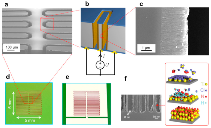
In-chip porous silicon-titanium nitride supercapacitor. (a) Scanning electron microscopy (SEM) inset of the trenches separating the electrodes (dark gray). (b) Rotated schematic illustration of the cross-section of two opposite electrodes of a device (titanium nitride-coated porous silicon layer with aluminum contact pads on the back side), with electrolyte shown in orange. (c) Higher-magnification SEM picture of the porous silicon regions. (d) Device trench side. (e) metallization side containing aluminum contacts for electrodes. (f) 3D illustration of two atomic-layer-deposition cycles of titanium-nitride growth. (credit: adapted from Kestutis Grigoras et al./Nano Energy)

Finnish researchers have developed a method for building highly efficient miniaturized micro-supercapacitor energy storage directly inside a silicon microcircuit chip, making it possible to power autonomous sensor networks, wearable electronics, and mobile internet-of-things (IoT) devices.

Supercapacitors function similar to standard batteries, but store electrostatic energy instead of chemical energy.

The researchers at VTT Technical Research Centre of Finland have developed a hybrid nano-electrode that’s only a few nanometers thick. It consists of porous silicon coated with a titanium nitride layer formed by atomic layer deposition.

The nano-electrode design features the highest-ever conductive surface-to-volume ratio. That combined with an ionic liquid (in a microchannel formed in between two electrodes), results in an extremely small form factor and efficient energy storage. That design makes it possible for a silicon-based micro-supercapacitor to achieve higher energy storage (energy density) and faster charge/discharge (power density) than the leading carbon- and graphene-based supercapacitors, according to the researchers.

The micro-supercapacitor can store 0.2 joule (55 microwatts of power for one hour) on a one-square-centimeter silicon chip. This design also leaves the surface of the chip available for active integrated microcircuits and sensors.

Micro-supercapacitors can also be integrated directly with active microelectronic devices to store electrical energy generated by thermal, light, and vibration energy harvesters to supply electrical energy (see, for example, Wireless device converts ‘lost’ microwave energy into electric power).

An open-access paper on the research has been published in Nano Energy journal.


Abstract of Conformal titanium nitride in a porous silicon matrix: A nanomaterial for in-chip supercapacitors

Today’s supercapacitor energy storages are typically discrete devices aimed for printed boards and power applications. The development of autonomous sensor networks and wearable electronics and the miniaturization of mobile devices would benefit substantially from solutions in which the energy storage is integrated with the active device. Nanostructures based on porous silicon (PS) provide a route towards integration due to the very high inherent surface area to volume ratio and compatibility with microelectronics fabrication processes. Unfortunately, pristine PS has limited wettability and poor chemical stability in electrolytes and the high resistance of the PS matrix severely limits the power efficiency. In this work, we demonstrate that excellent wettability and electro-chemical properties in aqueous and organic electrolytes can be obtained by coating the PS matrix with an ultra-thin layer of titanium nitride by atomic layer deposition. Our approach leads to very high specific capacitance (15 F cm−3), energy density (1.3 mWh cm−3), power density (up to 214 W cm−3) and excellent stability (more than 13,000 cycles). Furthermore, we show that the PS–TiN nanomaterial can be integrated inside a silicon chip monolithically by combining MEMS and nanofabrication techniques. This leads to realization of in-chip supercapacitor, i.e., it opens a new way to exploit the otherwise inactive volume of a silicon chip to store energy.

Hierarchies exist in the brain because of lower connection costs, research shows

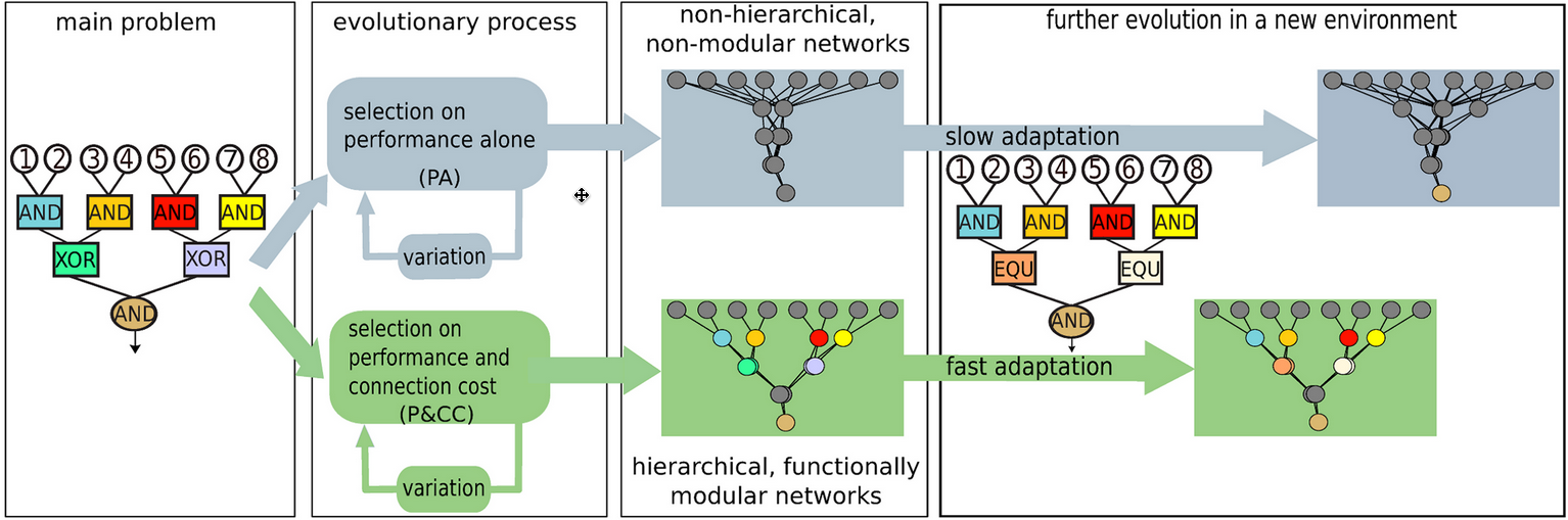
The Evolutionary Origins of Hierarchy: Evolution with performance-only selection results in non-hierarchical and non-modular networks, which take longer to adapt to new environments. However, evolving networks with a connection cost creates hierarchical and functionally modular networks that can solve the overall problem by recursively solving its sub-problems. These networks also adapt to new environments faster. (credit: Henok Mengistu et al./PLOS Comp. Bio)

New research suggests why the human brain and other biological networks exhibit a hierarchical structure, and the study may improve attempts to create artificial intelligence.

The study, by researchers from the University of Wyoming and the French Institute for Research in Computer Science and Automation (INRIA, in France), demonstrates that the evolution of hierarchy — a simple system of ranking — in biological networks may arise because of the costs associated with network connections.

This study also supports Ray Kurzweil’s theory of the hierarchical structure of the neocortex, presented in his 2012 book, How to Create a Mind.

The human brain has separate areas for vision, motor control, and tactile processing, for example, and each of these areas consist of sub-regions that govern different parts of the body.

Evolutionary pressure to reduce the number and cost of connections

The research findings suggest that hierarchy evolves not because it produces more efficient networks, but instead because hierarchically wired networks have fewer connections. That’s because connections in biological networks are expensive — they have to be built, maintained, etc. — so there’s an evolutionary pressure to reduce the number of connections.

In addition to shedding light on the emergence of hierarchy across the many domains in which it appears, these findings may also accelerate future research into evolving more complex, intelligent computational brains in the fields of artificial intelligence and robotics.

The research, led by Henok S. Mengistu, is described in an open-access paper in PLOS Computational Biology. The researchers also simulated the evolution of computational brain models, known as artificial neural networks, both with and without a cost for network connections. They found that hierarchical structures emerge much more frequently when a cost for connections is present.

Aside from explaining why biological networks are hierarchical, the research might also explain why many man-made systems such as the Internet and road systems are also hierarchical. “The next step is to harness and combine this knowledge to evolve large-scale, structurally organized networks in the hopes of creating better artificial intelligence and increasing our understanding of the evolution of animal intelligence, including our own,” according to the researchers.


Abstract of The Evolutionary Origins of Hierarchy

Hierarchical organization—the recursive composition of sub-modules—is ubiquitous in biological networks, including neural, metabolic, ecological, and genetic regulatory networks, and in human-made systems, such as large organizations and the Internet. To date, most research on hierarchy in networks has been limited to quantifying this property. However, an open, important question in evolutionary biology is why hierarchical organization evolves in the first place. It has recently been shown that modularity evolves because of the presence of a cost for network connections. Here we investigate whether such connection costs also tend to cause a hierarchical organization of such modules. In computational simulations, we find that networks without a connection cost do not evolve to be hierarchical, even when the task has a hierarchical structure. However, with a connection cost, networks evolve to be both modular and hierarchical, and these networks exhibit higher overall performance and evolvability (i.e. faster adaptation to new environments). Additional analyses confirm that hierarchy independently improves adaptability after controlling for modularity. Overall, our results suggest that the same force–the cost of connections–promotes the evolution of both hierarchy and modularity, and that these properties are important drivers of network performance and adaptability. In addition to shedding light on the emergence of hierarchy across the many domains in which it appears, these findings will also accelerate future research into evolving more complex, intelligent computational brains in the fields of artificial intelligence and robotics.

Electrical fields aid wound healing

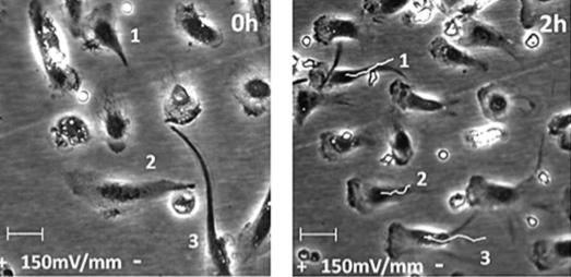
Human macrophages migrating directionally toward an electrode. Left: no electric field. Right: Time-lapse photo two hours after 150 mV/mm electric field applied (white lines shows the movement path toward candida yeast; numbers indicate start and end positions of cells). (credit: Joseph I. Hoare et al./JLB)

Small electrical currents appear to activate certain immune cells to jumpstart or speed wound healing and reduce infection when there’s a lack of immune cells available, such as with diabetes, University of Aberdeen (U.K.) scientists have found.

In a lab experiment, the scientists exposed healing macrophages (white blood cells that eat things that don’t belong), taken from human blood, to electrical fields of strength similar to that generated in injured skin. When the voltage was applied, the macrophages moved in a directed manner to Candida albicans fungus cells (representing damaged skin) to facilitate healing (engulfing and digesting extracellular particles). (This process is called “phagocytosis,” in which macrophages clean the wound site, limit infection, and  allow the repair process to proceed.)

The electric fields enhanced the uptake and clearance of a variety of targets known to promote inflammation and impair healing in addition to Candida albicans, including latex beads and expended white blood cells.*

“These findings raise the prospect that EF-based therapies could be extended beyond tissue repair and ultimately, be exploited to modulate the function of macrophages in other inflammatory diseases where these cells are dysregulated,” the researchers note in a report appearing in the June 2016 issue of the Journal of Leukocyte Biology.

“This new work identifies previously unappreciated opportunities to tune immune system function with electrical fields and has potentially wide-reaching implications for wound repair for a variety of diseases where macrophages play a role, including infectious disease, cancer and even obesity,” said John Wherry, Ph.D., Deputy Editor of the Journal of Leukocyte Biology.

The research extends previous research reported by KurzweilAI (New evidence that electrical stimulation accelerates wound healing).

* The experiments also showed that electric fields selectively augmented the production of protein modulators associated with the healing process, enhancing cytokine (growth factor) production and phagocytic activity essential for clearance of infection and for tissue repair and confirming that macrophages are tuned to respond to naturally generated electrical signals in a manner that boosts their healing ability.


Abstract of Electric fields are novel determinants of human macrophage functions

Macrophages are key cells in inflammation and repair, and their activity requires close regulation. The characterization of cues coordinating macrophage function has focused on biologic and soluble mediators, with little known about their responses to physical stimuli, such as the electrical fields that are generated naturally in injured tissue and which accelerate wound healing. To address this gap in understanding, we tested how properties of human monocyte-derived macrophages are regulated by applied electrical fields, similar in strengths to those established naturally. With the use of live-cell video microscopy, we show that macrophage migration is directed anodally by electrical fields as low as 5 mV/mm and is electrical field strength dependent, with effects peaking ∼300 mV/mm. Monocytes, as macrophage precursors, migrate in the opposite, cathodal direction. Strikingly, we show for the first time that electrical fields significantly enhance macrophage phagocytic uptake of a variety of targets, including carboxylate beads, apoptotic neutrophils, and the nominal opportunist pathogen Candida albicans, which engage different classes of surface receptors. These electrical field-induced functional changes are accompanied by clustering of phagocytic receptors, enhanced PI3K and ERK activation, mobilization of intracellular calcium, and actin polarization. Electrical fields also modulate cytokine production selectively and can augment some effects of conventional polarizing stimuli on cytokine secretion. Taken together, electrical signals have been identified as major contributors to the coordination and regulation of important human macrophage functions, including those essential for microbial clearance and healing. Our results open up a new area of research into effects of naturally occurring and clinically applied electrical fields in conditions where macrophage activity is critical.

How creating defective nanodiamonds could revolutionize nanotechnology and quantum computing

This electron microscope image shows a hybrid nanoparticle consisting of a nanodiamond (roughly 50 nanometers wide) covered in smaller silver nanoparticles that enhance the diamond’s optical properties. (credit: Min Ouyang)

University of Maryland researchers have developed a method to quickly and inexpensively assemble diamond-based hybrid nanoparticles from the ground up in large quantities while avoiding many of the problems with current methods.

These hybrid nanoparticles could speed the design of room-temperature qubits for quantum computers and create brighter dyes for biomedical imaging or highly sensitive magnetic and temperature sensors, for example.

When impurities are better

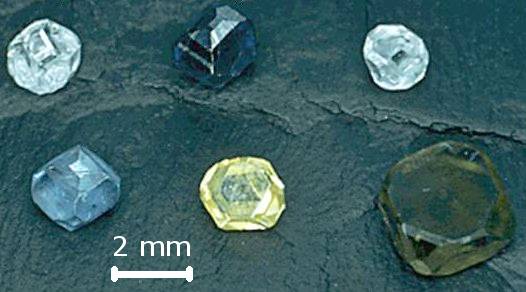
Synthetic diamonds of various colors (from defects) grown by the high-pressure high-temperature technique (credit: Wikipedia/
public domain)

The basic trick in creating a interesting or useful diamond is, ironically: Add a defect in the diamond’s crystal lattice. It’s similar to doping silicon to give it special electronic properties (such as making it work as a transistor).

Pure diamonds consist of an orderly lattice of carbon atoms and are completely transparent. However, pure diamonds are quite rare in natural diamond deposits; most have defects resulting from non-carbon impurities such as nitrogen, boron and phosphorus. Such defects create the subtle and desirable color variations seen in gemstone diamonds.

This altered bond is also the source of the optical, electromagnetic, and quantum physical properties that will make a nanodiamond useful when paired with other nanomaterials.

Nitrogen vacancy impurity

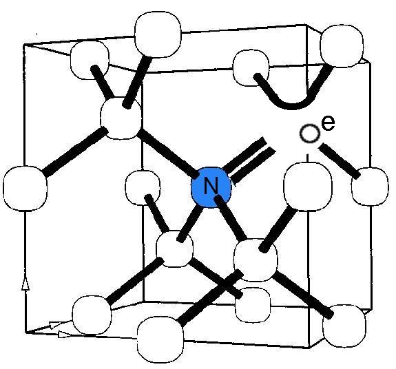
Model of nitrogen-vacancy center in diamond (credit: Wikipedia/public domain)

The most useful impurity — and used in the Maryland study — is the famous “nitrogen vacancy” defect: Sticking in a single nitrogen atom where a carbon atom should be, with an empty space right next to it.

As KurzweilAI has shown in several articles, a nitrogen vacancy in a diamond (or other crystalline materials) can lead to a variety of interesting new properties, such as a highly sensitive way to detect neural signals, an ultrasensitive real-time magnetic field detector, and importantly, making a nanodiamond behave as a quantum bit (qubit) for use in quantum computing and other applications.

Nearly all qubits studied to date require ultra-cold temperatures to function properly. A qubit that works at room temperature would represent a significant step forward, helping use quantum circuits in industrial, commercial and consumer-level electronics. That’s of special interest to Ougang’s team.

Volume production of hybrid nanoparticles

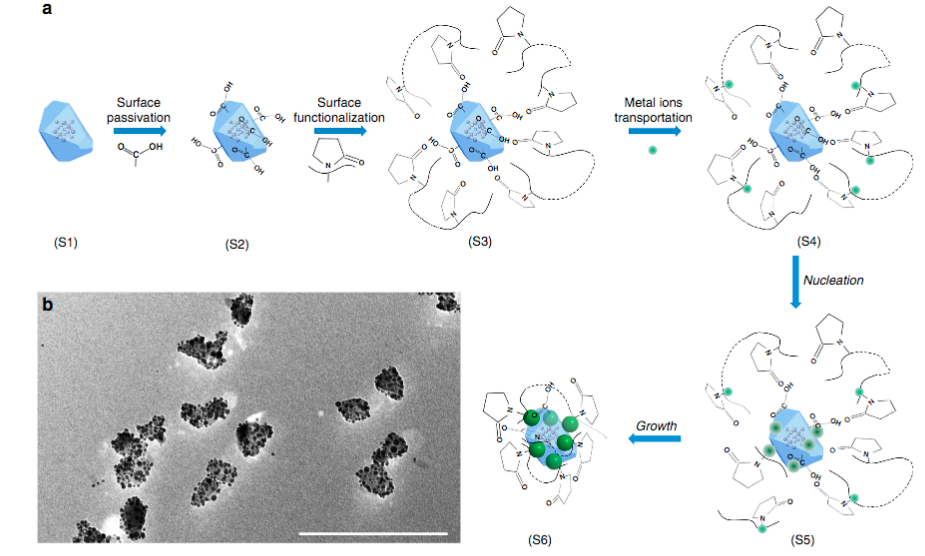
A synthetic route for hybrid nanodiamond nanoparticles. (a) Different growth stages, ending in (S6) growth of metal nanoparticles on the nanodiamond surface. (b) Transmission electron microscope image showing hybrid nanodiamond-silver nanostructures made by following the synthetic scheme in (a). Scale bar, 200 nm. (credit: J. Gong et al./Nature Communications)

Ougang’s and colleagues’ main breakthrough, though, is their method for constructing the hybrid nanoparticles. Other researchers have paired nanodiamonds with complementary nanoparticles using relatively imprecise methods, such as manually installing the diamonds and particles next to each other onto a larger surface one by one.

These top-down methods are costly, time consuming, and introduce a host of complications. “Our key innovation is that we can now reliably and efficiently produce these freestanding hybrid particles in large numbers,” explained Ouyang, who also has appointments in the UMD Center for Nanophysics and Advanced Materials and the Maryland NanoCenter, with an affiliate professorship in the UMD Department of Materials Science and Engineering.

His team’s method also enables precise control of the hybrid particles’ properties, such as the composition and total number of non-diamond particles.

“A major strength of our technique is that it is broadly useful and can be applied to a variety of diamond types and paired with a variety of other nanomaterials,” Ouyang said. “It can also be scaled up fairly easily. We are interested in studying the basic physics further, but also moving toward specific applications.”


Abstract of Nanodiamond-based nanostructures for coupling nitrogen-vacancy centres to metal nanoparticles and semiconductor quantum dots

The ability to control the interaction between nitrogen-vacancy centres in diamond and photonic and/or broadband plasmonic nanostructures is crucial for the development of solid-state quantum devices with optimum performance. However, existing methods typically employ top-down fabrication, which restrict scalable and feasible manipulation of nitrogen-vacancy centres. Here, we develop a general bottom-up approach to fabricate an emerging class of freestanding nanodiamond-based hybrid nanostructures with external functional units of either plasmonic nanoparticles or excitonic quantum dots. Precise control of the structural parameters (including size, composition, coverage and spacing of the external functional units) is achieved, representing a pre-requisite for exploring the underlying physics. Fine tuning of the emission characteristics through structural regulation is demonstrated by performing single-particle optical studies. This study opens a rich toolbox to tailor properties of quantum emitters, which can facilitate design guidelines for devices based on nitrogen-vacancy centres that use these freestanding hybrid nanostructures as building blocks.

Universe’s first life might have been born on diamond planets

In this artist’s conception, a carbon planet orbits a sunlike star in the early universe. Young planetary systems lacking heavy chemical elements but relatively rich in carbon could form worlds made of graphite, carbides, and diamond rather than Earth-like silicate rocks. Blue patches show where water has pooled on the planet’s surface, forming potential habitats for alien life. (credit: Christine Pulliam (CfA), Sun image: NASA/SDO)

New findings by scientists at the Harvard-Smithsonian Center for Astrophysics (CfA) suggest that planet formation in the early universe might have created carbon planets consisting of graphite, carbides, and diamond and that astronomers might find these diamond worlds by searching a rare class of stars.

“This work shows that even stars with a tiny fraction of the carbon in our solar system can host planets,” says lead author and Harvard University graduate student Natalie Mashian. “We have good reason to believe that alien life will be carbon-based, like life on Earth, so this also bodes well for the possibility of life in the early universe.”

The primordial universe consisted mostly of hydrogen and helium, and lacked chemical elements like carbon and oxygen necessary for life as we know it. Only after the first stars exploded as supernovae and seeded the second generation did planet formation and life become possible.

Clues to how life got started in the universe

Mashian and her PhD thesis advisor Avi Loeb examined a particular class of old stars known as carbon-enhanced metal-poor (CEMP) stars. These “anemic” stars contain only one hundred-thousandth as much iron as our Sun, meaning they formed before interstellar space had been widely seeded with heavy elements.

“These stars are fossils from the young universe,” explains Loeb. “By studying them, we can look at how planets, and possibly life in the universe, got started.”

CEMP stars have more carbon than would be expected, given their age. This relative abundance would influence planet formation as fluffy carbon dust grains (from supernovae) clump together to form tar-black worlds.

From a distance, these carbon planets would be difficult to tell apart from more Earth-like worlds. Their masses and physical sizes would be similar. Astronomers would have to examine their atmospheres for signs of their true nature. Gases like carbon monoxide and methane would envelop these unusual worlds.

The transit technique for detecting carbon planets

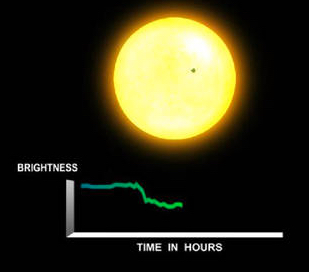
When a planet crosses in front of its star as viewed by an observer, the event is called a transit. The relative change in flux caused by a carbon-based planet transiting across its host CEMP star would range from ~0.0001% to ~0.01%.
(credit: NASA Ames)

But a dedicated search for planets around CEMP stars can be done using the transit technique, the scientists suggest. We encountered the transit technique on KurzweilAI in “How to use laser cloaking to hide Earth from remote detection by aliens” — in which we noted we two Columbia University astronomers suggested that we could cloak our Earth from aliens by shining a laser during transits so they couldn’t see the tiny giveaway changes in brightness — or that we could modify the light from our Sun during a transit to make it obviously artificial, sending a message to ET: “we’re here.”

In the new CfA paper, published in the Monthly Notices of the Royal Astronomical Society*, the scientists note that the relative change in flux caused by a carbon-based planet transiting across its host CEMP star ranges from ~0.0001% to ~0.01% — too weak to be detected by ground telescopes. That means it would require “space-based transit surveys that continuously monitor a large number of potential host stars over several years and measure their respective transit light curves.”

Fortunately, “there are a number of ongoing, planned, and proposed space missions committed to this cause,” the CfA scientists note. No word if the space-based transit surveys will also watch for artificial messages.

Headquartered in Cambridge, Mass., the Harvard-Smithsonian Center for Astrophysics (CfA) is a joint collaboration between the Smithsonian Astrophysical Observatory and the Harvard College Observatory. CfA scientists, organized into six research divisions, study the origin, evolution and ultimate fate of the universe.

* The CfA study is also available (open-access) on arXiv.


Abstract of CEMP stars: possible hosts to carbon planets in the early universe

We explore the possibility of planet formation in the carbon-rich protoplanetary disks of carbon-enhanced metal-poor (CEMP) stars, possible relics of the early Universe. The chemically anomalous abundance patterns ([C/Fe] ≥ 0.7) in this subset of low-mass stars suggest pollution by primordial core-collapsing supernovae (SNe) ejecta that are particularly rich in carbon dust grains. By comparing the dust-settling timescale in the protoplanetary disks of CEMP stars to the expected disk lifetime (assuming dissipation via photoevaporation), we determine the maximum distance rmax from the host CEMP star at which carbon-rich planetesimal formation is possible, as a function of the host star’s [C/H] abundance. We then use our linear relation between rmax and [C/H], along with the theoretical mass-radius relation derived for a solid, pure carbon planet, to characterize potential planetary transits across host CEMP stars. Given that the related transits are detectable with current and upcoming space-based transit surveys, we suggest initiating an observational program to search for carbon planets around CEMP stars in hopes of shedding light on the question of how early planetary systems may have formed after the Big Bang.

Deep neural networks to help identify, formulate advanced antiaging supplements

(credit: InSilico Medicine)

Insilico Medicine and Life Extension announced today an exclusive collaboration to identify novel biomarkers of human aging through the use of big-data analytics and AI, with the ultimate goal of discovery and formulation of nutrients to support health and longevity.

Insilico Medicine* is a big-data analytics company specializing in applying advances in deep learning to discovery of biomarkers and drugs. Life Extension**, a Florida-based organization established in the early 1980s, is a dietary-supplement innovator dedicated to extending healthy human longevity.

Insilico Medicine will focus on applying advanced signaling pathway activation analysis techniques and deep-learning algorithms to find nutraceuticals that mimic the tissue-specific transcriptional response of many known interventions and pathways associated with health and longevity.

Life Extension will use this information to develop novel nutraceutical products to support health and longevity, such as “geroprotectors” — precision natural organic small-molecule formulations that slow down or even reverse age-associated conditions and damage.

“Senescence is accompanied by a shift in cellular signaling that initiates and promotes a system-wide degenerative condition,” William Faloon, co-founder of the Life Extension Foundation, told KurzweilAI. “By turning back ‘on’ youthful signaling pathways while suppressing those that emerge during normal maturation, we expect to gain significant control over what are now thought to be inevitable pathologies of old age.”

“This collaboration means a lot to our team. For many years I was buying a range of supplements from Life Extension, and saw how passionate they are about extending healthy human life and supporting cutting-edge research in the field,” said Alex Zhavoronkov, PhD, CEO of Insilico Medicine, Inc. and the chief science officer of the Biogerontology Research Foundation in the UK. “Life Extension’s discovery research and product development teams have decades of experience in the pharmaceutical and nutraceutical research and have set very high standards for science-based nutraceutical discovery.”

Insilico Medicine scientists will present the results of this collaboration at RAADFest in San Diego in August, a longevity-focused conference.

Applying deep-learning techniques to aging

Insilico Medicine scientists have recently authored key papers on applying deep-learning techniques to biomedical applications in influential peer-reviewed journals, including “Developing criteria for evaluation of geroprotectors as a key stage toward translation to the clinic,” published in Aging Cell.

Other papers include “Deep learning applications for predicting pharmacological properties of drugs and drug repurposing using transcriptomic data” in Molecular Pharmaceutics, a journal published by the American Chemical Society; “Applications of Deep Learning in Biomedicine“, also in Molecular Pharmaceutics; and “Deep biomarkers of human aging: Application of deep neural networks to biomarker development” in Aging, a high-impact journal in aging research.

“We believe that accelerating the rate of progress in novel biomarker identification, as well as precision nutrient formulation development, is critical,” said Andrew G. Swick, Ph.D., senior vice president of scientific affairs, discovery research and product development for Life Extension.

“Life Extension aims to improve health and human longevity by utilizing the experience of noted research scientists and engaging in academic and industry collaborations. Artificial intelligence is one of the promising areas, where years of experience in the nutraceutical industry combined with sophisticated biologically inspired algorithms and high-performance computing may produce unprecedented results.”

* Insilico Medicine, Inc. is a bioinformatics company located at the Emerging Technology Centers at the Johns Hopkins University Eastern campus in Baltimore, with R&D resources in Belgium, Russia, and Poland. It utilizes advances in genomics, big-data analysis, and deep learning for in silico drug discovery and drug repurposing for aging and age-related diseases. The company pursues internal drug discovery programs in cancer, Parkinson’s, Alzheimer’s, sarcopenia, and geroprotector discovery. Through its Pharma.AI division, the company provides advanced machine-learning services to biotechnology, pharmaceutical, and skin-care companies.

** A trailblazer in the $35 billion U.S. dietary supplement industry for the past 36 years, Life Extension’s core mission is to extend the healthy human life span using an integrative approach by reporting on and funding cutting-edge scientific research. Life Extension Buyers Club (Life Extension) offers a full-range of premium-quality vitamins, minerals, and hormones as well as unique, specially made formulas. The company’s products are developed based on the latest scientific studies from peer-reviewed medical journals and are continually updated as new information occurs.